
| Table of Contents |  |
|
Case Report
| ||||||
| Management of an 11-week cesarean scar pregnancy | ||||||
| Sara Rocha1, Ana Carocha1, Catarina Marques1, Ana Gonçalves Andrade1, Álvaro Cohen1, Carlos Barros1 | ||||||
|
1Department of Obstetrics and Gynecology, Maternidade Dr. Alfredo da Costa - Centro Hospitalar Lisboa Central, Lisbon, Portugal.
| ||||||
| ||||||
|
[HTML Abstract]
[PDF Full Text]
[Print This Article]
[Similar article in Pumed] [Similar article in Google Scholar] |


| How to cite this article: |
| Rocha S, Carocha A, Marques C, Andrade AG, Cohen Á, Barros C. Management of an 11-week cesarean scar pregnancy. J Case Rep Images Gynecol Obstet 2015;1:10–14. |
|
Abstract
|
|
Introduction:
Cesarean scar pregnancy is a life-threatening condition, especially in the advanced gestational age. Optimal management has not yet been established because this is a rare condition.
Case Report: A 30-year-old female, with a previous cesarean, diagnosed with a viable cesarean scar pregnancy with 11 weeks of gestation. Successful termination of cesarean scar pregnancy was achieved with systemic and local (ultrasound-guided) methotrexate administration with subsequent dilatation and evacuation, under ultrasound guidance, in a two- stage procedure: rupture of the amniotic sac with partial removal of the fetus and one week later total evacuation of uterine content, with satisfactory hemostasis. Conclusion: Cesarean scar pregnancy is associated with a high morbidity and adverse consequences for future fertility. There is no optimal management established, because of the rarity of the condition. We present a case of successful termination of an advanced first trimester cesarean scar pregnancy. | |
|
Keywords:
Cesarean scar pregnancy, Intracardiac methotrexate injection, Uterine evacuation
| |
|
Introduction
| ||||||
|
Natural history of cesarean scar pregnancy (CSP) is unknown, but uterine rupture causing severe life-threatening bleeding, even in the first trimester, seems likely if the pregnancy is allowed to continue, with the possible need for hysterectomy and loss of fertility [1] [2]. According to available literature, there is no consensus for the treatment of CSP, especially in more advanced pregnancies. A variety of surgical and non-surgical interventions have been proposed for the treatment of CSP, but the best approach is still unknown. | ||||||
|
Case Report
| ||||||
|
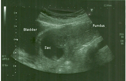
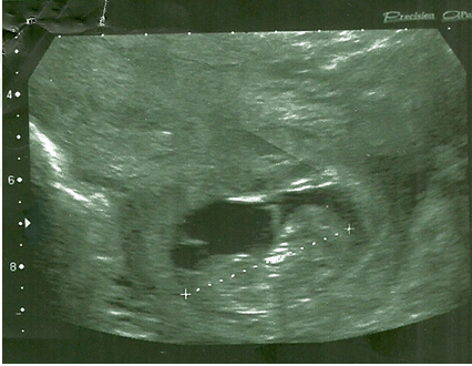
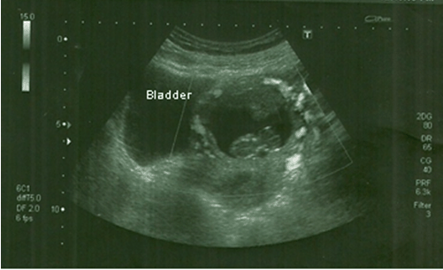
A 30-year-old female, gravid 4, para 2 (one cesarean due to fetal distress eleven years before, two spontaneous abortions with no uterine evacuation and 1 vaginal birth seven years before), was admitted to the emergency room with a history of a minor painless vaginal bleeding lasting for 14 days. Her period was in delay and she had a positive pregnancy test. Vaginal inspection revealed a cervix with normal appearance and a minor bleeding through a closed external os of the cervix. Ultrasound evaluation revealed an empty uterine cavity, a low implantation of the gestational sac, above a closed internal cervix (Figure 1) and a single fetus with cardiac activity and with a crown-rump length (40.8 mm) consistent with 10+5 weeks of gestation (Figure 2). The color Doppler study demonstrated increased flow in the anterior wall of the cervicoisthmic region and there was an absence of healthy myometrium between the bladder and the gestational sac (Figure 3). The initial human chorionic gonadotropin (β-hCG) concentration was 23,317 mIU/ml. Thus, ultrasound and analytic findings confirmed the diagnosis of a CSP. Methods of pregnancy termination were discussed with the patient. She chose medical treatment in order to preserve fertility. A total dose of 50 mg/m2 of body surface (85 mg) was divided between systemic methotrexate (MTX) and abdominal ultrasound-guided fetal intracardiac injection of MTX administered through a 20 G needle. The fetus heart activity ceased immediately. The procedure was uneventful. Day-4 quantitative β-hCG level was 18,635 mIU/ml and day-7 was 11,660 mIU/ml. Despite the favorable decline of β-hCG level, a second dose of 50 mg/m2 of body surface systemic MTX (85 mg) was administered at day-7. No adverse effects of MTX were noted. Initial determinations of β-hCG level kept showing decline (5961 mIU/ml and 1729 mIU/ml at day-10 and day-13, respectively), serial transvaginal ultrasound showed a gestational sac with no signs of detachment, a fetus with no cardiac activity and the color Doppler revealed no activity on the trophoblast area. The patient was discharged from the infirmary two weeks after admission and the follow-up treatment was conducted in an outpatient department with biweekly clinical, analytic (β-hCG monitoring) and ultrasound evaluation. Seven weeks after diagnosis the patient remained asymptomatic, the quantitative β-hCG level was 17 mIU/ml but ultrasound evaluation still showed a persistence of the gestational sac with no signs of abruption. For professional reasons, the patient wanted the resolution of her condition, so surgical treatment by ultrasound-guided transcervical curettage in a two-stage procedure was proposed to minimize the hemorrhagic risk. In the operating room, the patient was submitted to a partial uterine evacuation with rupture of the amniotic sac and partial removal of the fetus. There was a mild blood loss (less than 200 ml). One week later, β-hCG level was negative and ultrasound evaluation showed a collapsed gestational sac and partial detachment of trophoblast, so total evacuation of uterine content by cautious blunt curettage was performed. The procedure was successful with complete removal of the CSP, while maintaining satisfactory hemostasis (total blood volume loss of less than 500 ml). | ||||||
| ||||||
| ||||||
| ||||||
|
Discussion
| ||||||
|
Cesarean scar pregnancy must be distinguished from cervicoisthmic implantation, the latter sometimes resulting in term delivery [3]. In our case, ultrasonography revealed an empty uterine cavity, development of the gestational sac in the anterior part of the uterine isthmus, evidence of functional trophoblastic circulation on Doppler examination and an absence of healthy myometrium between the bladder and the gestational sac, allowing differentiation from cervicoisthmic implantation. Accordingly, based on the criteria postulated by Godin et al., the diagnosis of a CSP was made [4]. No consistent observational studies are available to create a universal treatment guideline for the management of CSP. A recent review exposed no less than 31 primary approaches to treat 751 cases of CSP and 44.1% ended up with complications. In this report, local (transvaginal or transabdominal ultrasound-guided) MTX injection, with or without additional intramuscular MTX administration, had the lowest complication rate (9.6%) [5]. Routes of MTX administration are systemically (normally by intramuscular injection) or locally: directly on the gestational sac (intragestacional) or on fetal heart (intracardiac). Available literature recommends against systemic MTX as a single treatment, because it could take days to stop fetal cardiac activity leading to additional growth of the embryo/fetus and vascularization of the sac, which may endanger the patient. This could be explained by the less vascularized fibrous tissue surrounding the scar section and the short half-life of MTX, which can limit local action of MTX. Local intra-gestational injection of MTX appears to be effective due to the high concentration of the drug in the sac. In the Timor-Tritsch et al. retrospective study of 26 cases of CSP, 19 were successfully treated with local and systemic MTX, including one case with 10 weeks of gestation and β-hCG level of 101000 mIU/ml [6]. Other authors also recommend local intra-gestational injections of MTX if β-hCG level of >10.00 mIU/ml, gestation sac diameter >2.5 cm and especially in the presence of embryonic/fetal cardiac activity [7]. Overall, combined systemic and local MTX have display good results and is well tolerated by the patient [7]. Initial management selected was combined systemic MTX and intracardiac MTX injection Because our patient had an advanced first trimester CSP. We chose intracardiac instead of intra-gestational MTX injection because, by directly entering fetal blood stream, trophoblastic blood flow can be more rapidly reduced and, therefore, hemorrhagic risk decreases. To our knowledge, this is the first case report that uses intracardiac MTX injection to treat a CSP. A consistent and favorable decrease of β-hCG level followed this procedure. Nevertheless, the patient remained hospitalized for two weeks and a second dose of systemic MTX was administered on day-7, firstly because our clinical experience in CSP is limited, and secondly because absolute value of β-hCG level was still elevated and we believed that the risk of complications was still high. As reported by some authors, it can take 4–16 weeks until the normalization of β-hCG levels and several months to a year until resolution of the CSP [8]. Accordingly, in our case, the normalization of β-hCG levels took eight weeks and curettage was necessary to remove the gestational sac. Dilatation and curettage has been applied as primary CSP treatment. A recent study from China reported 21 cases of CSP successfully treated with suction curettage alone, with no complications, if the myometrium layer between the gestational sac and the bladder had more than 3 mm and before 10 gestational weeks [9]. Other authors presented suction curettage as a viable alternative for conservative treatment in patients with CSP diagnosed before eight gestational weeks and who have a myometrium thickness of more than 4.5 mm [10] . In another study from China, 30 from 100 patients underwent ultrasound-guided curettage with no complications [11]. Nevertheless, suction curettage alone for CSP is still controversial. Complications include uterine perforation, massive bleeding and need for hysterectomy [12]. It is more consensual to perform ultrasound-guided curettage when the serum β-hCG returns to near normal levels, no trophoblastic blood flow is detected and a connection between the gestational sac and the uterine cavity is noted on the ultrasound [7]. Other authors advise that correct timing of curettage after MTX intervention is when the mass is <4 cm in diameter without profuse peritrophoblastic flow, β-hCG is <2000 mIU/L and myometrium layer between the bladder and the sac is >4 mm [13]. In a recent study, local and systemic MTX administration followed by suction curettage, followed by Foley tamponade, was an effective treatment in treating 42 of the 45 patients with CSP [14]. Also, combined MTX plus curettage treatment results in a shorter time of therapy, can prevent massive hemorrhage occurring acutely in an outpatient setting and can avoid unnecessary laparotomy [7]. In our case, to minimize the hemorrhagic risk, transcervical procedure guided by abdominal ultrasound was performed by an experienced doctor in a two-stage procedure already described. The goal was to allow a gradual detachment of the trophoblast and facilitate its removal in a second procedure. To our knowledge, there is no other study that reports the same approach. Nevertheless, potential risks like local hematoma, uterine scar rupture, bladder injury and life-threatening hemorrhage must be taken into account. | ||||||
|
Conclusion
| ||||||
|
At the moment there are no universal treatment guidelines for the management of cesarean scar pregnancy (CSP), especially in more advanced pregnancies. To standardize treatment modalities, all cases of CSP should be reported and further multicentric studies are also needed. Combined systemic and local (preferably intracardiac) methotrexate injection is a good option for women who want to preserve fertility and cautious uterine evacuation can abbreviate the resolution of CSP. These are treatments that, globally, most Maternity Hospitals are able to perform, but potential risks must be taken into account and doctors must be prepared for emergency intervention. | ||||||
|
References
| ||||||
| ||||||

|
[HTML Abstract]
[PDF Full Text]
|
|
Author Contributions
Sara Rocha – Substantial contributions to conception and design, Acquisition of data, Analysis and interpretation of data, Drafting the article, Final approval of the version to be published Ana Carocha – Acquisition of data, Analysis and interpretation of data, Drafting the article, Final approval of the version to be published Catarina Marques – Acquisition of data, Analysis and interpretation of data, Drafting the article, Final approval of the version to be published Ana Gonçalves Andrade – Substantial contributions to conception and design, Drafting the article, Final approval of the version to be published Álvaro Cohen – Substantial contributions to conception and design, Revising it critically for important intellectual content, Final approval of the version to be published Carlos Barros – Substantial contributions to conception and design, Revising it critically for important intellectual content, Final approval of the version to be published |
|
Guarantor of submission
The corresponding author is the guarantor of submission. |
|
Source of support
None |
|
Conflict of interest
Authors declare no conflict of interest. |
|
Copyright
© 2015 Sara Rocha et al. This article is distributed under the terms of Creative Commons Attribution License which permits unrestricted use, distribution and reproduction in any medium provided the original author(s) and original publisher are properly credited. Please see the copyright policy on the journal website for more information. |
|
|