
 |
|
Case Report
| ||||||
| Ovarian metastasis of gastrointestinal stromal tumor: A case report and review of the literatures | ||||||
| Mayumi Kobayashi1, Hiroshi Yoshida2, Reiko Watanabe2, Tomoyasu Kato4 | ||||||
|
1MD, Division of Gynecology, National Cancer Center Hospital, Tokyo, Japan
2MD, PhD, Departments of Pathology and Clinical Laboratories, National Cancer Center Hospital, Tokyo, Japan | ||||||
| ||||||
|
[HTML Abstract]
[PDF Full Text]
[Print This Article]
[Similar article in Pumed] [Similar article in Google Scholar] |


| How to cite this article: |
| Kobayashi M, Yoshida H, Watanabe R, Kato T. Ovarian metastasis of gastrointestinal stromal tumor: A case report and review of the literatures. J Case Rep Images Gynecol Obstet 2017;3:9–12. |
|
ABSTRACT
|
|
Introduction:
Ovarian metastases of gastrointestinal stromal tumors (GISTs) are extremely rare, however, these patients may still have a favorable prognosis when treated with imatinib after resection.
Case Report: We report on the successful treatment of a 54-year-old female with gastric GIST metastatic to the ovary, who had undergone distal gastrectomy 11 years ago. The resected ovarian tumor was a solid multinodular mass consisting of KIT-positive and discovered on gastrointestinal stromal tumors protein (DOG1)-positive spindle cells, and was thus diagnosed as a recurrent metastasis of gastric GIST. No tumor recurrence was observed during the two years of follow-up while on imatinib therapy. Conclusion: As GIST metastatic to the ovary may mimic primary ovarian tumors, KIT and DOG1 immunohistochemistry can aid in making correct pathological diagnosis. Furthermore, patients with this particular metastatic pattern may benefit from imatinib treatment after resection. | |
|
Keywords:
Gastrointestinal stromal tumor, Kitt, Metastasis, Ovary
| |
|
INTRODUCTION
| ||||||
|
Gastrointestinal stromal tumors (GISTs) are potentially malignant and are the most common mesenchymal tumors of the gastrointestinal tract. They often spread to extra-intestinal sites. However, ovarian metastasis of GISTs is extremely rare. There have been less than 10 reported cases treated by surgical resection [1][2] [3][4], therefore information on clinical experience of this unusual situation is very limited. As GISTs often demonstrate various morphologies, they can mimic primary ovarian tumors. Immunohistochemical staining for KIT and DOG1 can be utilized to provide a definitive diagnosis. We report a successfully treated case of ovarian metastasis from a gastric GIST that presented 11 years after the first surgical resection. | ||||||
|
CASE REPORT
| ||||||
|
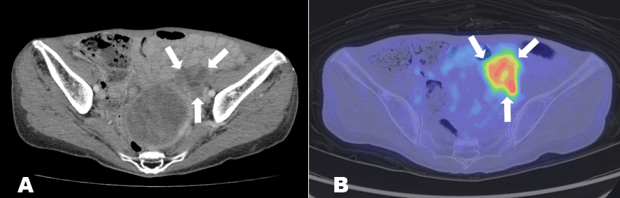
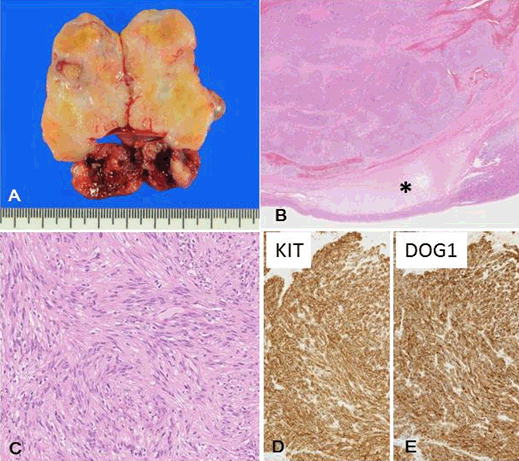
A 54-year-old female underwent distal gastrectomy for the resection of a primary gastric GIST at her previous hospital. Three years after the operation, a recurrence of GIST under her left diaphragm along with the spleen was detected and surgically removed. Three years later, she had another tumor recurrence in the remnant stomach and liver. Following the resection, she received imatinib (400 mg/day per os), a tyrosine kinase inhibitor, for two months. After the patient experienced severe side effects from imatinib, specifically edema and diarrhea, the dose was decreased to 200 mg and subsequently discontinued after four months. At this time, she was referred to our hospital. Five years after her last operation (11 years after the first operation), a 30-mm mass was detected in the lower pole of the left kidney on a computed tomography (CT) scan. An 18-fluoro-deoxyglucose-positron emission tomography (18FDG-PET) scan revealed an area of increased uptake of 18FDG in a pelvic lesion adjacent to the intestinal tract that was discrete from the left renal mass (Figure 1). On transvaginal ultrasonography, no abnormalities were observed except for a uterine leiomyoma. Upon undergoing her fourth operation for the removal of recurrent masses, intra-operative findings revealed tumors located under the left diaphragm, in the lower pole of the left kidney, and on the left ovary. A total abdominal hysterectomy with bilateral salpingo-oophorectomy was performed in addition to tumor resections at the left diaphragm and left kidney. Macroscopically, the tumor in the left ovary was a yellow-to-gray-colored solid multinodular mass. Microscopically, the tumor consisted of fascicles of uniform, spindle-shaped tumor cells with elongated nuclei and increased chromatin. The tumor cells had strong and diffuse immunoreactivity for KIT and DOG1. The tumor was then diagnosed as an ovarian metastasis from the primary gastric GIST (Figure 2). Imatinib treatment was restarted at a lower dose of 200 mg/day. Although mild edema developed, she was able to tolerate continuing imatinib therapy with a gradual dose increase from 200–300 mg/day. No additional side effects were reported and no tumor recurrences were detected during the two years of follow-up. | ||||||
| ||||||
|
| ||||||
| ||||||
|
DISCUSSION
| ||||||
|
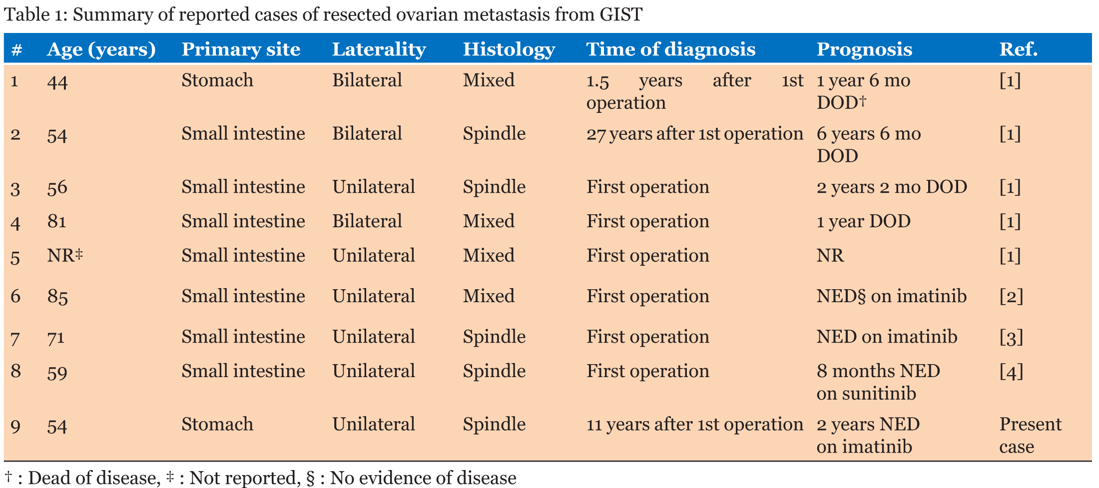
We reported a case of gastric GIST metastatic to the ovary treated with surgical resection and adjuvant imatinib therapy. Despite this unusual metastatic pattern, her detailed medical history and the immunohistochemistry of the tumor aided us in making a correct diagnosis. Ovarian metastasis of GISTs is extremely rare, with only eight case reports in English and Japanese literatures (Table 1). Ovarian metastases were discovered with the initial diagnosis in six cases [1] [2][3][4] and as recurrences in two cases [1]. GISTs may originate from the interstitial cells of Cajal or their stem cell-like precursors and can occur anywhere along the gastrointestinal tract, but are most often found in the stomach (60%) or small intestine (20%) [5]. However, 87.5% of tumor metastases to the ovaries originated in the small bowel. GISTs of small bowel origin are known to be more likely to recur or metastasize than gastric GISTs [1]. The present case is evidence that gastric GISTs may metastasize to the ovary after a latency period of 11 years. To the best of our knowledge, this is the first report in which ovarian metastasis of a gastric GIST has been successfully treated using imatinib therapy following surgical resection. The malignant potential of GISTs ranges from small lesions with no recurrent risk to aggressive sarcomas. The prognosis of a patient with primary GIST depends on tumor size, location and mitotic activity [6] . The overall prognosis of a patient with GIST confined to the primary organ is favorable and the five-year relative survival was reported to be 91%. On the other hand, the five-year relative survival was 48% for the patients with metastatic GIST [7]. Before the imatinib era, median survival of patients with recurrent or metastatic GIST was 10–20 months, but after the introduction of imatinib and other tyrosine kinase inhibitors, median survival of advanced GIST was reported to be 51–57 months [7]. The prognosis of patients with ovarian metastasis from GISTs seems to have improved dramatically with the advent of imatinib treatment. Irving et al. reported death in all cases of ovarian metastases in which imatinib was not administered, with the exception of one case that was lost to follow-up [1]. However, there have been no reported cases of death following adjuvant imatinib and/or sunitinib therapy. Therefore, at the present time, the correct diagnosis of this unusual metastatic pattern may change patients’ prognoses. The diversity of GIST morphology generates various differentials. GISTs can demonstrate spindle cell, epithelioid cell, or mixed cell patterns. In regards to the ovarian metastases of the eight cases previously reported, four cases showed spindle cell patterns and the rest had mixed cell patterns. Thus, the differential diagnosis includes various types of primary ovarian tumors such as fibromas, the comas, smooth muscle tumors, endometrial stromal tumors, schwannomas, adult granulosa cell tumors, and metastatic carcinomas [1]. It is difficult to differentiate metastatic GISTs from these primary ovarian tumors based only on morphology. However, a detailed medical history and immunohistochemistry can help make the correct diagnosis and, subsequently, determine the optimal therapy. KIT and DOG1, discovered on GIST 1, are now regarded as the most useful immunohistochemical markers of GISTs. Most GISTs have gain-of-function mutations of the KIT or platelet-derived growth factor receptor (PDGFRA) genes [8]. Immunohistochemically, 90-95% of GISTs are KIT-positive tumors that have membranous and/or cytoplasmic staining patterns. However, GISTs harboring PDGFRA mutations are more likely to express KIT-negativity. Moreover, as a potential pitfall, KIT-positive tumors that are not GISTs have been reported, such as undifferentiated gastric carcinoma [9]. DOG1 has emerged in recent years as a promising biomarker for GISTs. There is a small subset of KIT-negative GISTs that are positive for DOG1. Similar immunoreactivity is rarely observed in other mesenchymal and non-mesenchymal tumor types [10]. DOG1 was also reported to have a high sensitivity of more than 95% for GISTs with a high mitotic index and/or increased nuclear pleomorphism [11]. Our case showed typical spindle cell morphology and concomitant KIT and DOG1 immunoreactivity, which led us to the correct diagnosis of gastric GIST metastatic to the ovary. | ||||||
|
CONCLUSION
| ||||||
|
In conclusion, we reported a successfully treated case of gastric GIST metastatic to the ovary 11 years after the initial operation. Immunohistochemistry for KIT and DOG1 can be useful for making a definitive diagnosis of ovarian metastasis of a GIST. Furthermore, with the advent of imatinib therapy, it is possible that even patients with ovarian metastasis of GISTs may show a favorable prognosis if the correct diagnosis is made and subsequent optimal treatment is implemented. | ||||||
|
REFERENCES
| ||||||
| ||||||

|
[HTML Abstract]
[PDF Full Text]
|
|
Acknowledgements
We acknowledge Editage’s editorial support in preparing this manuscript. |
|
Author Contributions
Mayumi Kobayashi – Substantial contributions to conception and design, Acquisition of data, Analysis and interpretation of data, Drafting the article, Revising it critically for important intellectual content, Final approval of the version to be published Hiroshi Yoshida – Substantial contributions to conception and design, Acquisition of data, Analysis and interpretation of data, Drafting the article, Revising it critically for important intellectual content, Final approval of the version to be published Reiko Watanabe – Substantial contributions to conception and design, Drafting the article, Revising it critically for important intellectual content, Final approval of the version to be published Tomoyasu Kato – Acquisition of data, Analysis and interpretation of data, Drafting the article, Revising it critically for important intellectual content, Final approval of the version to be published |
|
Guarantor of submission
The corresponding author is the guarantor of submission. |
|
Source of support
None |
|
Conflict of interest
Authors declare no conflict of interest. |
|
Copyright
© 2017 Mayumi Kobayashi et al. This article is distributed under the terms of Creative Commons Attribution License which permits unrestricted use, distribution and reproduction in any medium provided the original author(s) and original publisher are properly credited. Please see the copyright policy on the journal website for more information. |
|
|