 |
Case Report
Rare association of exomphalos and maternal alcohol use
1 MBChB, Gold Coast University Hospital, University of Queensland, Queensland, Australia
2 MBBCh, FRCOG, FRANZCOG, Toowoomba Base Hospital, University of Queensland, Queensland, Australia
Address correspondence to:
Sabiha Mohamad Zakaria
MBChB, Gold Coast University Hospital, Queensland,
Australia
Message to Corresponding Author
Article ID: 100131Z08SM2022
Access full text article on other devices

Access PDF of article on other devices


How to cite this article
Mohamad Zakaria S, Kassab A. Rare association of exomphalos and maternal alcohol use. J Case Rep Images Obstet Gynecol 2022;8(2):34–37.ABSTRACT
Introduction: Omphalocele is a congenital anomaly characterized by an abdominal wall defect. Chromosomal, syndromic, or other structural anomalies often co-exist and largely affect the prognosis.
Case Report: We discuss a case of a complex omphalocele associated with thoracic hypoplasia and thoracic-lumbar scoliosis in a chromosomally unaffected fetus.
Conclusion: This is an unusual case where this congenital defect is potentially associated to maternal alcohol use or even herpes simplex virus (HSV).
Keywords: Abdominal wall defect, Alcohol, Congenital HSV, Omphalocele
Introduction
Abdominal wall defect is a common congenital anomaly, usually diagnosed prenatally on ultrasound. Omphalocele is a serious form of this and is characterized by abdominal contents (bowel and/or abdominal viscera) in a sac outside the abdomen through a midline abdominal wall defect, with the umbilicus inserting into the base of the sac. Size can vary from small to very large. A giant omphalocele is usually more than 5 cm in size and holds 75% of the fetal liver outside the abdomen. Fetal outcome depends on the defect size and the severity of any associated structural or chromosomal anomaly—which occur in about half of the cases. Trisomy 18 makes up for about 75% of chromosomally abnormal cases, while Beckwith–Wiedemann syndrome, found in 4% of omphaloceles, is the most common syndromic association.
Case Report
The mother is a 29-year-old in her 7th pregnancy with three live births (aged 16, five, and one), two terminations of pregnancy, and one miscarriage. The first pregnancy was largely uneventful and term delivery was via an emergency lower segment caesarean section for fetal distress. Birth weight was 4.6 kg. In the two subsequent vaginal deliveries, the second child required a 2-day Neonatal Intensive Care stay for respiratory complications, and the third child was well but small for gestational age. She had a history of depression and mild asthma and was not on regular medications. She reported a history of treated chlamydia eight years ago but no genital herpes. She is of indigenous origin. She smoked 15 cigarettes a day. Alcohol use out of pregnancy was about 7–15 units/week which she reduced significantly to minimal sporadic usage on discovering her pregnancy at about eight weeks gestation. Her body mass index was 25.
She presented to her general practitioner (GP) at 11 weeks. She was then not on folate. Her blood group was AB positive and antibody screen was negative. Hepatitis, human immunodeficiency virus (HIV), syphilis, chlamydia, and gonorrhea screens were normal. She was non-immune to rubella. HB1Ac was 5.0. Her first trimester screen was calculated low risk but an omphalocele was noted. Nuchal translucency was 2.8 mm. She was therefore referred to a Maternal Fetal Medicine Department.
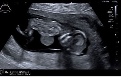
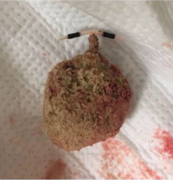
Her tertiary ultrasound undertaken at 14 weeks showed a large abdominal wall defect with a mass protruding through the upper abdominal wall. However, the lesion was complex: The cord inserted into the right of the mass and the umbilical vein took an irregular course confirming that the liver was within the mass. A large amount of bowel and possibly the stomach were extruded outside the abdominal cavity. No membrane covered the mass. Thoracolumbar scoliosis and thoracic hypoplasia were noted. No anomalies of the heart, bladder, amniotic fluid, arms, legs, and neck were noted (Figure 1, Figure 2, Figure 3).
The patient was counseled regarding risk of possible co-existent chromosomal or structural anomalies, intrauterine growth restriction, fetal hydrops, likely tertiary hospital caesarean delivery, prolonged neonatal hospital stay and prognosis. The couple opted for a medical termination of pregnancy without any invasive testing which was uneventful at 15 weeks.
Contemporaneous maternal bloods tests including viral serology as well as a medical panel were unremarkable. Herpes simplex virus immunoglobulin G (IgG) serology was not done at the time. Fetal karyotype was 46XY and chromosomal microarray showed no clinically significant copy number changes. The placenta was immature, weighted <5th centile and demonstrated established post-intrauterine death changes. Swabs cultured normal regional flora. Microbiological polymerase chain reaction (PCR) was undertaken on the placental tissue including syphilis, toxoplasmosis, Q fever, parvovirus, cytomegalovirus, and HSV. All were negative except HSV 2 PCR which came back positive.


Discussion
At the detection of the complex omphalocele, a chromosomal anomaly or Pentalogy of Cantrell was suspected. However, a normal karyotype and positive placental HSV-2 PCR raise the possibility of a viral etiology.
Herpes simplex virus exists as two subtypes: HSV-1 and HSV-2—which occur independently of each other. It is extremely common: HSV-1 affects 67% of the population by the age of 50. 1HSV-1 is mainly transmitted by oral-to-oral contact to cause oral herpes, in the form of painful ulcers, while both can cause genital disease. The infection can be asymptomatic and is lifelong, undergoing intermittent reactivation especially if triggered by immunosuppression, stress, and illness. Severe HSV is usually seen in the immunosuppressed as meningoencephalitis and disseminated infection. Transmission is highest with active blisters but can occur through asymptomatic individuals. Diagnosis is by a positive PCR swab of the lesion. A negative HSV-specific IgG serology undertaken concurrently implies a primary infection, while a positive result indicates a secondary infection.
Herpes simplex virus in pregnancy is clinically important. The mother is at increased risk of recurrence especially in the third trimester. Maternal disseminated infection is rare. Neonatal manifestation can be limited to SEM (skin, eyes, mucous membrane) or the central nervous system or disseminated disease with multi organ failure. The latter two are associated with significant mortality and neurological morbidity. Fortunately, neonatal infections are rare: 10 in 100,000 cases globally [1]. 90% of the transmission occurs during passage through an infected birth canal and the risk of transmission is highest (50%) if the infection is acquired in the third trimester or if not seroconverted despite an earlier infection. In such cases, caesarean section delivery is usually indicated for neonatal safety.
Congenital HSV infections are extremely rare and generally present as cerebral anomalies and intrauterine growth restriction on prenatal ultrasound. A literature review of cases of congenital HSV on PubMed between 1989 and 2017 by Fa et al. describes 36 cases of congenital HSV, of which only 10 were diagnosed prenatally. These were overwhelmingly a variety of cerebral anomalies, and the less common non-cerebral anomalies related to skin, eyes, heart, liver, spleen, and limbs [2]. Similar findings are noted in the literature review by Marquez et al. [3]. Abdominal wall defects do not figure in these literature reviews, as well as the authors’ own PubMed search of congenital HSV anomalies. Interestingly 48% and 43% of the mothers were asymptomatic of a HSV infection in these two reviews, as was our patient. Notwithstanding, Finger-Jardim et al.’s report on HSV PCR tests on 422 placentas demonstrated that the prevalence of HSV-1 and HSV-2 was, respectively, 28% and 12.6% (maternal side) and 29.9% and 8.3% (fetal side)—figures much higher than the incidence of congenital HSV [4]. This could suggest that placental HSV-positivity is a common occurrence and does not necessarily translate to fetal infection.
Another possible teratogen at embryogenesis is maternal alcohol. Fetal alcohol spectrum disorders are characterized by specific craniofacial anomalies, neurodevelopmental disorders, growth restriction, and cardiac anomalies, but traditionally not omphalocele. A retrospective observational study of 27,438 births at a German University hospital showed that none of the 18 cases of omphalocele over an 11-year period was associated with maternal alcohol [5]. However in the National Birth Defect Prevention Study undertaken across multiple US centers, the adjusted Odd Ratio between periconceptional maternal alcohol consumption and the 254 cases of chromosomally normal and non-syndromic omphalocele was 1.50 (CI 1.15–1.96). 46% of the mothers affected by an omphalocele reported alcohol intake in the pregnancy in this cohort [6]. Interestingly, smoking has no statistically significant influence on the incidence of omphalocele [5].
Thoracic hypoplasia—seen in our case—can be associated with skeletal dysplasias such as Saldino–Noonan syndrome and Beemer–Langer syndrome. Cardiac, genitourinary and limb anomalies often co-exist, but concurrent omphalocele, HSV infection, or alcohol use is not a described association of this anomaly.
Conclusion
While the definite etiology of this complex omphalocele remains elusive, both HSV and alcohol are extremely rare possible explanations and should be considered in its etiopathogenesis after ruling out common causes.
REFERENCES
1.
Herpes simplex virus. 2022. [Available at: https://www.who.int/news-room/fact-sheets/detail/herpes-simplex-virus]

2.
Fa F, Laup L, Mandelbrot L, Sibiude J, Picone O. Fetal and neonatal abnormalities due to congenital herpes simplex virus infection: A literature review. Prenat Diagn 2020;40(4):408–14. [CrossRef]
[Pubmed]
3.
Marquez L, Levy ML, Munoz FM, Palazzi DL. A report of three cases and review of intrauterine herpes simplex virus infection. Pediatr Infect Dis J 2011;30(2):153–7. [CrossRef]
[Pubmed]
4.
Finger-Jardim F, Avila EC, da Hora VP, Gonçalves CV, de Martinez AMB, Soares MA. Prevalence of herpes simplex virus types 1 and 2 at maternal and fetal sides of the placenta in asymptomatic pregnant women. Am J Reprod Immunol 2017;78(1). [CrossRef]
[Pubmed]
5.
Kapapa M, Rieg, T, Henne-Bruns D, Serra A. Risk factors for abdominal wall defects. Congenital Anomalies 2020;60(2):54–61. [CrossRef]
6.
Richardson S, Browne ML, Rasmussen SA, et al. Associations between periconceptional alcohol consumption and craniosynostosis, omphalocele, and gastroschisis. Birth Defects Res A Clin Mol Teratol 2011;91(7):623–30. [CrossRef]
[Pubmed]
SUPPORTING INFORMATION
Author Contributions
Sabiha Mohamad Zakaria - Conception of the work, Design of the work, Acquisition of data, Analysis of data, Drafting the work, Revising the work critically for important intellectual content, Final approval of the version to be published, Agree to be accountable for all aspects of the work in ensuring that questions related to the accuracy or integrity of any part of the work are appropriately investigated and resolved.
Ahmed Kassab - Conception of the work, Design of the work, Revising the work critically for important intellectual content, Final approval of the version to be published, Agree to be accountable for all aspects of the work in ensuring that questions related to the accuracy or integrity of any part of the work are appropriately investigated and resolved.
Guaranter of SubmissionThe corresponding author is the guarantor of submission.
Source of SupportNone
Consent StatementWritten informed consent was obtained from the patient for publication of this article.
Data AvailabilityAll relevant data are within the paper and its Supporting Information files.
Conflict of InterestAuthors declare no conflict of interest.
Copyright© 2022 Sabiha Mohamad Zakaria et al. This article is distributed under the terms of Creative Commons Attribution License which permits unrestricted use, distribution and reproduction in any medium provided the original author(s) and original publisher are properly credited. Please see the copyright policy on the journal website for more information.


