| Table of Contents | |
|
Case Report
| ||||||
| Uterine arteriovenous malformation inadvertently treated by hysteroscopy resection | ||||||
| Giancarlo Garuti1, Attilio Di Spiezio Sardo3, Maurizio Mirra2, Stefania Calabrese3, Cristina Gonfiantini2, Marco Di Mario1 | ||||||
|
1Obstetric and Gynecology Department, Public Hospital of Lodi, via Savoia 1, 26900-Lodi, Italy.
2Pathology Department, Public Hospital of Lodi, via Savoia 1, 26900-Lodi, Italy. 3Department of Gynecology and Obstetrics and Pathology of Human Reproduction, University of Naples "Federico II°", via Pansini 5, Naples, Italy. | ||||||
| ||||||
|
[HTML Abstract]
[PDF Full Text]
[Print This Article]
[Similar article in Pumed] [Similar article in Google Scholar] |
| How to cite this article: |
| Garuti G, Sardo ADS, Mirra M, Calabrese S, Gonfiantini C, Di Mario M. Uterine arteriovenous malformation inadvertently treated by hysteroscopy resection. J Case Rep Images Gynecol Obstet 2015;1:20–24. |
|
Abstract
|
|
Introduction:
Uterine arteriovenous malformations (AVMs) are one of the causes of potential severe genital hemorrhages. Acquired AVMs are considered iatrogenic and mainly diagnosed after pregnancy termination and/or uterine surgery. The diagnosis is based on ultrasound, computed tomography, magnetic resonance imaging and angiography. Uterine artery embolization and hysterectomy represent the current treatments of choice. Uterine curettage is not recommended for AVMs treatment due to high risk of hemorrhage. Although hysteroscopy is the reference method for studying intra-uterine pathologies, few reports described hysteroscopy features of AVMs. We report on a patient with an AVM occasionally diagnosed and managed by hysteroscopy, a treatment never reported in literature.
Case Report: A 52-year-old patient complaining of abnormal uterine bleeding, submitted to a cesarean section 29 years before, was scheduled to hysteroscopy resection of an intrauterine lesion suggestive of submucosal myoma at saline infusion ultrasonography. Rather than a myoma, hysteroscopy imaging was consistent with an endometrial polyp with no abnormal vascularization. The slicing of the mass was hampered by bleeding from a crowding of arterial and venous vessels; near the pedicle, a worsening of bleeding precluded an adequate visualization causing the premature interruption of the surgery. Bleeding control was obtained by the placement of a balloon. The pathologic examination confirmed an AVM. One month later, ultrasound, computed tomography and hysteroscopy showed no residual mass. Twelve months after intervention no vaginal bleeding was recorded and normal findings were found at physical and ultrasound examination. Conclusion: AVMs can be misdiagnosed as a submucous myoma or an endometrial polyp at ultrasounds and hysteroscopy, respectively. Although theoretically effective, hysteroscopy resection of AVMs can lead to hemorrhagic complications. | |
|
Keywords:
Abnormal uterine bleeding, Endometrium, Hysteroscopy, Uterine arteriovenous malformations
| |
|
Introduction
| ||||||
|
Uterine arteriovenous malformations (AVMs) are rare vascular lesions causing abnormal and possible life-threatening bleeding [1] [2]. They are classified as congenital, resulting from abnormality in the embryonic uterine angiogenesis and acquired, usually traumatic and resulting from prior uterine curettage, pregnancy termination, cesarean section or uterine surgery [3]. The common anatomical landmark is represented by a tangle of vessels showing connections between feeding arteries and draining veins without interposed capillary network [4]. Functionally, AVMs show high blood-flow and low resistance, presenting as color mosaic with flow reversal on Color Doppler Ultrasound (US) and low-impedance and high-velocity on spectral analysis [5]. Angiography computed tomography (CT) and magnetic resonance imaging (MRI) scan can define feeder vessels and the anatomy of AVMs [6]. Angiography is considered the gold standard for AVMs diagnosis, recognizing feeder arteries and early venous filling [3] [7]. Hysteroscopy is the reference technique to assess intrauterine pathology but currently there are few reports assessing the endoscopic imaging of AVMs [8][9][10][11]. To our knowledge in current literature no patients with AVM underwent hysteroscopy treatment. We report the case of a patient with a uterine AVM unintentionally treated by hysteroscopy, following a preoperative diagnosis of submucous myoma. | ||||||
|
Case Report
| ||||||
|
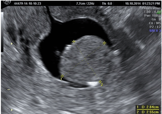
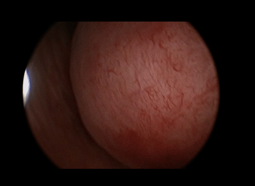
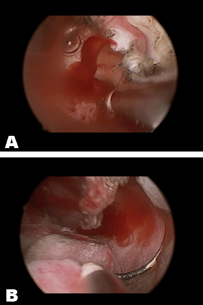
A 52-years-old female without significant medical history was scheduled for hysteroscopy resection of an intrauterine mass. She had an obstetrical history of cesarean section at 23-years-old due to breech presentation and an uncomplicated vaginal birth at 36-years-old. In the last four years chronic heavy periodic menstrual bleeding and intermenstrual bleeding have been reported. Oral estroprogestins have been administered during the last 18 months with no improvement of symptoms. In October 2014, transvaginal gray scale US detected an endometrial lesion measuring 28 mm in largest diameter, suspected to be a fundal submucous myoma by saline infusion sonography (Figure 1). No further investigation was accomplished. No abnormality was found at physical examination. Hysteroscopy was carried out under conscious sedation by an experienced surgeon (GG). After cervical dilatation, uterine cavity was assessed by a 27 Fr resectoscope fitted with 4 mm bipolar loop (Versapoint Bipolar System, Gynecare, Ethicon Inc., Menlo Park CA) set at 160 W power and Versapulse vapor-cutting mode. Continuous saline flow was delivered at working pressure set at 40 mmHg by an electronic irrigation-suction device. Hysteroscopy identified a soft mass sizing 30 mm, arising from the uterine fundus by a large pedicle. The covering mucosal lining was evenly smooth, neither prominent vascularization nor pulsatile vessels were found. Based on these findings, rather than a myoma, inspective features oriented the surgeon to a polyp-like mass (Figure 2). Since the beginning of resection, a heavy bleeding was originated from crowded vessels showing both arterial (pulsating, roundly-shaped vessels showing a thickened muscular wall) and venous (non pulsating, oval shaped vessels showing a thin wall) features (Figure 3A-B). An extensive use of low-frequency current was applied to achieve hemostasis. The mass was almost entirely removed but near its pedicle an uncontrollable bleeding hampered the safe visualization of surgical field. The procedure was stopped and a Foley catheter was inserted into the endometrial cavity and 10 cc of saline was inflated to control the blood loss. The surgery lasted 23 minutes without any saline deficit recorded. After three hours from intervention the balloon was removed, no significant bleeding was observed and the patient was discharged after two hours of clinical monitoring. The pathologic assessment reported the following: "Vascular malformation formed by arterial and venous vessels dissociating smooth muscle fibers" (Figure 4). In order to evaluate vascularization and extent of the residual uterine mass, one month after intervention the patient was re-assessed by eco Color Doppler Transvaginal US and TC angiography. Both examinations did not show evidence of any mass or abnormal vascularity. Two months after intervention a second-look hysteroscopy confirmed the absence of residual mass and only an uneven thickening of fibrous consistence were found instead of pedicle. A coagulation of this endometrial area was accomplished. After 12 months from surgery no abnormal bleeding was recorded and no abnormalities were found at physical examination and transvaginal US. | ||||||
| ||||||
|
| ||||||
| ||||||
| ||||||
|
Discussion
| ||||||
|
Hysteroscopy imaging of uterine AVM has been described as normal appearing endometrium [11], pulsatile vessels bulging of the endometrial cavity [10] [12]and more frequently as highly vascular masses indistinguishable from Retained Products of Conception (RPOC) [8][9] [13]. On color Doppler US, RPOC [14], accreta [15] and subinvolution of the placental bed [5] share features common to AVMs, due to the high-flow and low-resistance characterizing placental blood flow [16]. In the majority of the reports, the diagnosis of AVMs was given only through US after termination of pregnancy and in several series an uneventful, spontaneous and fast resolution of AVM was detected [1] [3][5][7][17][18]. Therefore, several AVMs reported in literature were probably pregnancy-related complications instead of real structural vascular abnormalities [3] [7][16][18]. Far from pregnancy termination or in menopausal patients, in absence of the bias related to the persistence of a misleading placental blood-flow, AVMs are rarely reported [1][2][4][6][19]. Pathologic findings on uterine specimens or endometrial biopsies are rarely recorded as support of AVMs diagnosis [4] [12][13][17], due to either the expanding use of Uterine Artery Embolization (UAE) as primary therapy or to the belief that endometrial surgical instrumentation can worsen uterine bleeding [2][3] [7][14]. The treatment options for AVMs depend on clinical presentation. In asymptomatic patients or in case of non severe uterine bleeding, expectant management and medical hormone therapies or methylergometrine have shown to be effective. In anemic or hemodynamically unstable patients selective UAE is the preferred treatment of AVMs, especially in women who desire to preserve fertility. Other conservative surgical managements (less frequently reported) are laparotomy removal, laparoscopic bipolar coagulation and ligation of the uterine artery. Hysterectomy is suggested in cases of life threatening bleeding or when fertility preservation is not of concern [3]. To our knowledge, hysteroscopy resection was never reported for AVMs treatment. In the above presented patient, a diagnosis of AVM, was pathologically confirmed and it was probably originally related to past cesarean section. The US imaging detected an intrauterine mass mimicking a submucous myoma and showed polyp-like features at hysteroscopy-view. Before intervention, gray-scale US underestimated the vascular nature of the endometrial mass and color Doppler assessment was not applied. The electrosurgical resection of the lesion caused an unexpected excessive bleeding, poorly controlled by coagulating current and hampering its safe surgical removal even under hysteroscopy vision. In this case, the bleeding was stopped by an endometrial compressive device and the AVM was entirely removed by a single-step hysteroscopy surgery. This positive result was probably related to the favorable topography of AVM itself, mainly expanding within endometrial cavity and sparing myometrium. | ||||||
|
Conclusion
| ||||||
|
Uterine arteriovenous malformations (AVMs) can present myoma-like or polyp-like features at ultrasound scan and hysteroscopy assessment, respectively. A color Doppler investigation should be always considered when the clinical background cannot safely exclude the above reported rare (but potentially life-threatening) cause of uterine hemorrhage. The experience presented confirms the common belief that in cases of suspected AVM, primary endometrial surgical instrumentation such as D&C and even hysteroscopy resection should be avoided or managed with extreme caution, due to the high risk of uterine hemorrhage. | ||||||
|
Acknowledgements
| ||||||
|
We thank Mrs Elena Bosoni for language revision of the Manuscript. | ||||||
|
References
| ||||||
| ||||||
|
[HTML Abstract]
[PDF Full Text]
|
|
Author Contributions
Giancarlo Garuti – Substantial contributions to conception and design, Acquisition of data, Analysis and interpretation of data, Drafting the article, Revising it critically for important intellectual content, Final approval of the version to be published Attilio Di Spiezio Sardo – Analysis and interpretation of data, Revising it critically for important intellectual content, Final approval of the version to be published Maurizio Mirra – Analysis and interpretation of data, Revising it critically for important intellectual content, Final approval of the version to be published Stefania Calabrese – Analysis and interpretation of data, Revising it critically for important intellectual content, Final approval of the version to be published Cristina Gonfiantini – Analysis and interpretation of data, Revising it critically for important intellectual content, Final approval of the version to be published Marco Di Mario – Analysis and interpretation of data, Revising it critically for important intellectual content, Final approval of the version to be published |
|
Guarantor of submission
The corresponding author is the guarantor of submission. |
|
Source of support
None |
|
Conflict of interest
Authors declare no conflict of interest. |
|
Copyright
© 2015 Giancarlo Garuti et al. This article is distributed under the terms of Creative Commons Attribution License which permits unrestricted use, distribution and reproduction in any medium provided the original author(s) and original publisher are properly credited. Please see the copyright policy on the journal website for more information. |
|
|