|
Case Report
MRI and placenta accreta: Keys for its interpretation in images, regarding a case
1 Radiology Resident, Facultad de Medicina Clínica Alemana de Santiago, Universidad del Desarrollo, Vitacura, Santiago, Chile
2 Radiologist, Fellowship in Abdominal Imaging, Departamento de imágenes abdominales, Clínica Alemana de Santiago, Vitacura, Santiago, Chile
3 Radiologist, Departamento de imágenes abdominales, Clínica Alemana de Santiago, Vitacura, Santiago, Chile
4 Gynecologist, Clínica Alemana de Santiago, Vitacura, Santiago, Chile
5 Pathologist, Clínica Alemana de Santiago, Vitacura, Santiago, Chile
Address correspondence to:
Francisca Aliaga
Avenida Vitacura 5951, Vitacura, Santiago,
Chile
Message to Corresponding Author
Article ID: 100186Z08FA2024
Access full text article on other devices

Access PDF of article on other devices

How to cite this article
Aliaga F, del Campo F, Cocio R, Schiappacasse G, Blanco S, Pires Y. MRI and placenta accreta: Keys for its interpretation in images, regarding a case. J Case Rep Images Obstet Gynecol 2024;10(2):50–53.ABSTRACT
Introduction: Placenta accreta spectrum (PAS) refers to the abnormal adherence of the placental trophoblast to the uterine myometrium. Several conditions are associated with its development. Ultrasound is the imaging modality of choice for antenatal diagnosis. In cases of doubt, magnetic resonance (MR) plays a leading role, due to its high performance, and contributes to surgical planning. The final diagnosis is made in the surgery, with subsequent confirmation by pathology. The objective of this article is to present a case of placenta increta, focused on the imaging findings in magnetic resonance imaging (MRI).
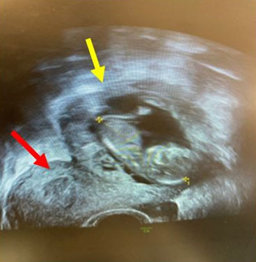
Case Report: We present a case of a 35-year-old woman with a history of 2 previous cesarean sections and chronic hypertension, who presented a 23-week pregnancy with obstetric ultrasound showing placenta accreta. An MRI was performed, and it showed signs of placenta accreta, with no evidence of transmural extension to adjacent organs. At 34 weeks, a cesarean section and hysterectomy were performed, with no evidence of bladder invasion.
Conclusion: The diagnosis of PAS is made antenatal with ultrasound, but MRI has been used in some cases, with some characteristic features that every radiologist should know.
Keywords: Hysterectomy, Magnetic resonance, Placenta accreta, Ultrasound
SUPPORTING INFORMATION
Acknowledgments
Departments of Radiology, Anatomical Pathology, Gynecology and Obstetrics, Facultad de Medicina Clínica Alemana, Universidad del Desarrollo, Santiago de Chile.
Author ContributionsFrancisca Aliaga - Substantial contributions to conception and design, Acquisition of data, Analysis of data, Interpretation of data, Drafting the article, Revising it critically for important intellectual content, Final approval of the version to be published
Fernanda del Campo - Drafting the article, Revising it critically for important intellectual content, Final approval of the version to be published
Rolando Cocio - Substantial contributions to conception and design, Acquisition of data, Analysis of data, Interpretation of data, Drafting the article, Revising it critically for important intellectual content, Final approval of the version to be published
Giancarlo Schiappacasse - Substantial contributions to conception and design, Revising it critically for important intellectual content, Final approval of the version to be published
Sebastián Blanco - Analysis of data, Interpretation of data, Revising it critically for important intellectual content, Final approval of the version to be published
Yumay Pires - Acquisition of data, Interpretation of data, Revising it critically for important intellectual content, Final approval of the version to be published
Guaranter of SubmissionThe corresponding author is the guarantor of submission.
Source of SupportNone
Consent StatementWritten informed consent was obtained from the patient for publication of this article.
Data AvailabilityAll relevant data are within the paper and its Supporting Information files.
Conflict of InterestAuthors declare no conflict of interest.
Copyright© 2024 Francisca Aliaga et al. This article is distributed under the terms of Creative Commons Attribution License which permits unrestricted use, distribution and reproduction in any medium provided the original author(s) and original publisher are properly credited. Please see the copyright policy on the journal website for more information.