|
Case Report
Neurofibromatosis type 2 diagnosed at time of cervical squamous cell carcinoma
1 Resident, Department of Gynecologic Surgery and Obstetrics, Tripler Army Medical Center, Tripler AMC, HI, USA
2 Gynecologic Oncologist, Department of Gynecologic Surgery and Obstetrics, Tripler Army Medical Center, Tripler AMC, HI, USA
Address correspondence to:
Emily Penick
MD, FACOG, 1 Jarrett White Road, Tripler AMC, HI 96859,
USA
Message to Corresponding Author
Article ID: 100242Z08CL2026
Access full text article on other devices

Access PDF of article on other devices

How to cite this article
Lester C, Penick E. Neurofibromatosis type 2 diagnosed at time of cervical squamous cell carcinoma. J Case Rep Images Obstet Gynecol 2026;12(2):37–41.ABSTRACT
Introduction: Cervical cancer is most frequently diagnosed in women between ages 35–44 and rarely develops in younger women. Over 90% of cervical cancers are associated with human papillomavirus (HPV) persistence, but some are associated with genetic mutations such as STK11. This case discusses development of squamous cell carcinoma in a 25-year-old patient following low-grade squamous intraepithelial lesion (LSIL), cannot exclude high grade cytology result four years prior.
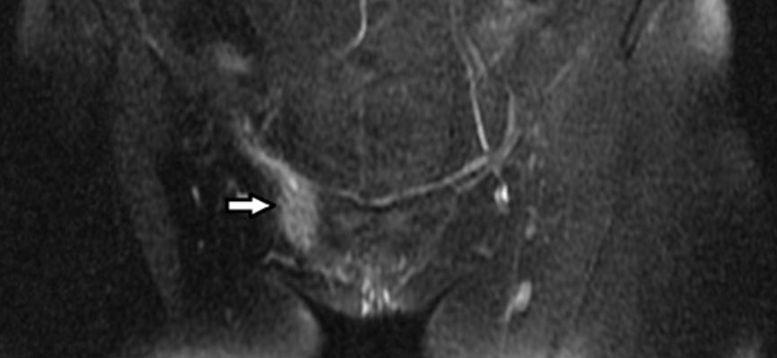
Case Report: A 25-year-old G2P2 presented for a postpartum exam, which was notable for a 1 cm lesion of the anterior cervix. Biopsies confirmed squamous cell carcinoma. A positron emission tomography/computed tomography (PET/CT) was completed for surgical planning which noted bilateral brain masses, concerning for vestibular schwannomas, which met clinical criteria for neurofibromatosis type 2 (NF2). Following referral to neurosurgery, a left retrosigmoid craniotomy was completed. A modified radical abdominal hysterectomy and sentinel lymph node dissection was performed following recovery from neurosurgery. Somatic genetic testing returned with a frameshift mutation in NF2E1194FJ. Final hysterectomy pathology noted a grade 3 tumor measuring 1.2 cm in size and with 3.75 mm depth of invasion.
Conclusion: Low-grade intraepithelial lesion cytology typically has a high probability of regression and low risk for progression to cancer. Neurofibromin 2 regulates many cellular events. Studies note aberrant expression of NF2 contributes to tumorigenesis in other cancers. The tumorigenesis of both HPV and NF2 may share synergistic mechanisms which lead to progression to carcinoma. While the clinical course of our patient exhibits both a clinical diagnosis of NF2 and somatic NF2 loss in her cervical tumor, we cannot definitively conclude that NF2 deficiency accelerated the timeline of her cervical carcinogenesis. Rather, this clinical scenario prompts an intriguing hypothesis regarding potential molecular synergy between high-risk HPV oncoproteins and loss of the NF2 gene product, Merlin.
Keywords: Cervical cancer, Human papillomavirus, Neurofibromatosis 2
SUPPORTING INFORMATION
Author Contributions
Clayton Lester - Substantial contributions to conception and design, Acquisition of data, Analysis of data, Interpretation of data, Drafting the article, Revising it critically for important intellectual content, Final approval of the version to be published
Emily Penick - Substantial contributions to conception and design, Acquisition of data, Analysis of data, Interpretation of data, Drafting the article, Revising it critically for important intellectual content, Final approval of the version to be published
Guaranter of SubmissionThe corresponding author is the guarantor of submission.
Source of SupportNone
Consent StatementWritten informed consent was obtained from the patient for publication of this article.
Data AvailabilityAll relevant data are within the paper and its Supporting Information files.
Conflict of InterestAuthors declare no conflict of interest.
Copyright© 2026 Clayton Lester et al. This article is distributed under the terms of Creative Commons Attribution License which permits unrestricted use, distribution and reproduction in any medium provided the original author(s) and original publisher are properly credited. Please see the copyright policy on the journal website for more information.