 |
Case Report
Spontaneous anterior abdominal wall expulsion of female sterilization Filshie clips
1 BA, MA, MBBS, Medical Officer, Department of Obstetrics and Gynaecology, Singapore General Hospital, Singapore
2 MD, MRCOG, Registrar, Department of Obstetrics and Gynaecology, Singapore General Hospital, Singapore
3 MBBS, MMed, MRCOG, FAMS, Senior Consultant, Department of Obstetrics and Gynaecology, Singapore General Hospital, Singapore
Address correspondence to:
Hui Men Selina Chin
Department of Obstetrics and Gynaecology, Singapore General Hospital, Outram Road, 169608,
Singapore
Message to Corresponding Author
Article ID: 100090Z08HC2021
Access full text article on other devices

Access PDF of article on other devices


How to cite this article
Chin HMS, He S, Lim SK. Spontaneous anterior abdominal wall expulsion of female sterilization Filshie clips. J Case Rep Images Obstet Gynecol 2021;7:100090Z08HC2021.ABSTRACT
Introduction: Filshie clip ligation is a common procedure for female sterilization. Rarely, Filshie clips may dislodge and migrate through tissue planes (0.6%) involving bladder, appendix, inguinal canal, vagina, urethra, and rectum. The pathophysiology is unclear. We discuss a case of delayed Filshie clip expulsion from the anterior abdominal wall.
Case Report: A 35-year-old Chinese lady presented with a few-month history of a 6 × 5 cm superficial, firm, tender, and mobile infra-umbilical lump with purulent discharge and surrounding erythema. She was afebrile and her inflammatory markers were not raised. Obstetrical history included three lower segment caesarean sections via Pfannenstiel incision, and Filshie clip postpartum sterilization three years ago. She denied other medical or surgical history. Computerized tomography showed a 2.1 cm hyper-dense soft tissue in the infra-umbilical anterior abdominal wall containing two ligation clips. She opted for conservative management with antibiotics. The clips expelled spontaneously from the abdominal lump in succession over the next month—both were closed and complete. Subsequent hysterosalpingography showed abrupt truncation in bilateral fallopian tubes with no peritoneal spillage.
Conclusion: Spontaneous anterior abdominal wall expulsion of Filshie clips is a rare complication of tubal ligation that may occur from weeks to years after. When patients are counseled pre-operatively, risks of clip migration and expulsion should be discussed. Sterilization history should also be sought in females presenting with abdominal pain.
Keywords: Expulsion, Filshie clip, Migration, Tubal ligation
Introduction
Sterilization is the most common form of female contraception worldwide. Commonly, Filshie clip ligation is preferred for tubal occlusion. Filshie clip is a silicone-lined titanium clip that acts as an occlusive device when applied over the fallopian tubes. Migration of Filshie clips has been well documented in literature. An additional complication of clip extrusion has recently received attention, with two large studies of tubal sterilization describing clip extrusion which collectively report six extrusions in 6707 women (0.09% incidence) [1]. Migration of Filshie clips through tissue planes has been described in literature to involve bladder, appendix, inguinal canal, vagina, urethra, and rectum [2], however the pathophysiology is unclear. Here, we discuss a case of delayed Filshie clip expulsion from the anterior abdominal wall.
Case Report
A 35-year-old Chinese lady presented to the Gynecology clinic with a few-month history of a spontaneous painful abdominal lump below her umbilicus. She denied any prior trauma, abdominal lumps, or skin infections. There were no urinary or bowel symptoms, nausea, or fever.
Her obstetrical history included three lower segment caesarean sections (LSCS) via Pfannenstiel incision, the last of which included postpartum sterilization using Filshie clips three years ago. There were no complications intra-operatively and no documented difficulty in the application of clips. Both fallopian tubes were identified by tracing to fimbrial ends before Filshie clip application. She denied any other medical or surgical history. Her menstrual history was unremarkable.
On physical examination, she was afebrile and her vital signs were normal. Abdominal examination revealed a 6 × 1 cm superficial, firm, mildly tender, mobile sub-umbilical lump. Cough impulse was negative. Her LSCS scar was well healed.
Abdominal wall ultrasonography revealed a 5.9 × 5.5 × 2.6 cm mixed solid cystic lesion within the subcutaneous layer and in close proximity to the superior aspect of the bladder. As it was in close proximity to the LSCS scar, impression was of an organized hematoma or previous partially resolved collection. Internal vascularity was noted within the solid component of the lesion, indicating early inflammatory change.
A week later, she presented to the Emergency Department with worsening pain, redness, and bruising over the lump. Physical examination revealed a 6 × 5 cm tender indurated lump with purulent discharge from a 4 cm central fluctuant area, with surrounding erythema and warmth. She remained afebrile and her vital signs were stable. Complete blood count and electrolytes were normal with a total white cell count of 5.5 × 10⁹/L and C-reactive protein (CRP) of 2.5 mg/L. Urine analysis and urine culture were unremarkable. She was treated for abdominal wall abscess with intravenous antibiotics. Swab culture taken from the discharge was negative for bacterial growth.
Pelvic ultrasonography revealed normal uterus and ovaries. No obvious abdominal wall endometriotic deposit or abdominal masses were identified.
Computerized tomography of her abdomen and pelvis showed a 2.1 cm hyperdense soft tissue in the infra-umbilical anterior abdominal wall, which was contiguous with the anterior wall of the urinary bladder. Within this thickening, two tubal ligation clips were seen. Small and large bowel loops appeared to be uninvolved (Figure 1).
In view of these findings, exploratory laparotomy was offered to remove the Filshie clips. She opted for conservative management with oral antibiotics.
A month later, she noticed extrusion of a Filshie clip from the site of discharge, which was subsequently removed. The abdominal mass had resolved with a 5 mm area of slight erythema.
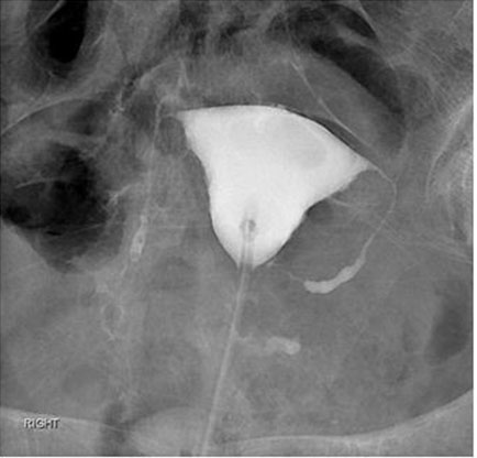
She was managed with oral antibiotics and daily wound dressings. Two weeks later, the second Filshie clip expelled spontaneously in a similar way. Both clips were examined to be closed and complete. She subsequently underwent hysterosalpingography which confirmed non-patency of bilateral fallopian tubes (Figure 2).

Discussion
Female sterilization is the most preferred contraception method worldwide, accounting for 30.2% in the United States. With failure rates of 0.5%, it is a safe and effective option for permanent contraception. Tubal ligation via laparoscopy or laparotomy utilizes a range of different techniques (complete or partial salpingectomy, bipolar coagulation, silicone band, or spring clip).
Tubal occlusion via Filshie clips or other mechanical occlusion methods are generally preferred over tubal electrocoagulation due to an avoidance of electrical burns from electrocoagulation, reduced risks of ectopic pregnancy, and lesser tubal damage which increases the chance of reversal by tubal anastomosis [2]. A Filshie clip applied to full thickness of fallopian tubes leads to local avascular necrosis, with the silicone component expanding to keep the lumen blocked. This is followed by division and healing of the stumps at both sides of the clip [3]. It is theorized that the clip is held in place by peritonealization and adhesion formation, thus if this process is slow to occur, clip detachment may occur. Dislodgement of Filshie clip from the fallopian tube into the peritoneal cavity is well-recognized, and is usually harmless and asymptomatic. The most common sites of Filshie clip migration are in the Pouch of Douglas or paracolic gutters [4]. Filshie clip expulsion has been reported to occur via anterior abdominal wall (4), perianal or pararectal tissues (4), groin (3), colon (2), vagina (3), and urinary bladder (2) [2],[4],[5],[6].
Our case is the fifth reported case worldwide of Filshie clip migration and expulsion via the anterior abdominal wall [7],[8],[9],[10]. A painful lump was noted in all five cases with discharge noted in three of them, and the location of abscess was mostly sub-umbilical.
Although immediate post-operative recovery occurs within months, it has been suggested that the remodeling phase of wound healing may extend for years beyond the primary procedure. A report of 54 patients presenting for tubal ligation reversal identified tubal patency in 17% of subjects at a mean of 4.8 years after tubal ligation, which was thought to be secondary to tuboperitoneal fistulae. This was in contrast to the short-term failure rate of tubal ligation at 1–5% at three months post-procedure. Thus, asymptomatic and undetected tuboperitoneal fistulae may occur more commonly than originally thought [5],[11].
Common presentations of Filshie clip migration include abscess formation, ulceration, fistula formation, and tissue induration, with resolution of symptoms after clip expulsion or retrieval, suggesting that symptoms are part of a local tissue inflammatory response. Absence of fever/leucocytosis and negative bacterial cultures indicate against true infection and abscess formation. Time of presentation also varies greatly—from six weeks up to 21 years after application [12].
Interestingly, there appears to be no risk factor that predisposes one to clip migration. In majority of cases, pelvic anatomy was observed to be normal without pathology or intra-operative complications during laparoscopic ligation. There was no apparent association between site of migration, history of gynecological procedures or pelvic pathology, and duration to presentation.
Although it remains a possibility, it is unlikely that Filshie clips were possibly applied incorrectly in this case, due to the normal pelvic anatomy, lack of intra-operative complications, complete documentation that the tubes were identified correctly, complete occlusion noted on subsequent hysterosalpingography, and the fact that the clips were closed upon visual inspection after expulsion.
It has also been proposed that clip expulsion may in part be due to an immune-mediated reaction to foreign materials [5]. Filshie clips are made of titanium, and are silicone-lined. Silicone has been thought to be relatively inert, hence its extensive use as coating agents. However, cases of silicone as an allergenic material causing implant expulsion have been reported, including in cochlear implants and pacemakers, where resolution of symptoms depended on subsequent implantation of non-silicone-based devices [13],[14]. Type four hypersensitivity is characterized by localized abundance of macrophages and T lymphocytes, with an absence of B lymphocytes on histological examination [15], however this is also seen in autoimmune responses and certain pathogenic infections. Skin patch testing for allergy may be considered, but a negative result does not necessarily eliminate contact allergy as a diagnosis. While it may be tempting to attribute Filshie clip extrusion to an allergenic reaction in this case, it is also notable that there are reported cases where the second Filshie clip was retained with symptom resolution after expulsion of only one clip via the anterior abdominal wall [7]. If an allergenic reaction was suspected, both clips would probably have been expelled.
Diagnosis of clip migration and subsequent expulsion is often revealed only after radiological imaging, with common presentations being abscess formation, ulceration, fistula formation, and tissue induration. Hence, the physician should be mindful to consider clip migration in his differential diagnosis of abdominal pain in the absence of obvious pathology, and proactively enquire regarding obstetric and gynecological history, including any previous sterilization.
Although Filshie clip extrusion has not been associated with a significant risk of harm or long-term adverse sequelae, it may be a matter of medicolegal importance. This unexpected complication also understandably produces anxiety in patients who may perceive the event as a failure of sterilization, and perhaps it may be worth mentioning during pre-operative counseling. Other alternatives of permanent female contraceptive methods such as bilateral salpingectomy should be discussed and offered in addition to tubal ligation, with the benefit of reducing the overall risk of epithelial ovarian cancers.
Conclusion
Filshie clip extrusion is a rare complication, however in the small collection of published in case reports, it is clear that they may migrate transperitoneally in the pelvic cavity and between tissue planes to be expulsed from the abdominal wall, among other distal sites. This may occur between weeks and years after initial placement. When patients are counseled pre-operatively, risks of clip migration and expulsion should be discussed. Sterilization history should also be sought in females presenting with abdominal pain to improve clinical diagnosis.
REFERENCES
1.
Sokal D, Gates D, Amatya R, Dominik R. Two randomized controlled trials comparing the tubal ring and filshie clip for tubal sterilization. Fertil Steril 2000;74(3):525–33. [CrossRef]
[Pubmed]

2.
Gad N, Aziz R, Siwicki K. Filshie clip migration into wall of urinary bladder presenting with acute abdominal pain. Case report and review of English literature: From 1990 to April 2009. Pelviperineology 2010;29:84–7.
3.
Filshie GM, Casey D, Pogmore JR, Dutton AG, Symonds EM, Peake AB. The titanium/silicone rubber clip for female sterilization. Br J Obstet Gynaecol 1981;88(6):655–62. [CrossRef]
[Pubmed]
4.
Kesby GJ, Korda AR. Migration of a Filshie clip into the urinary bladder seven years after laparoscopic sterilisation. Br J Obstet Gynaecol 1997;104(3):379–82. [CrossRef]
[Pubmed]
5.
Fahey M. Spontaneous expulsion of tubal ligation clips: A case report. J Obstet Gynaecol Can 2007;29(9):733–6. [CrossRef]
[Pubmed]
6.
Kale A, Chong YS. Spontaneous vaginal expulsion of a Filshie clip. Ann Acad Med Singap 2008;37(5):438–9.
[Pubmed]
7.
Lok IH, Lo KWK, Ng JSW, Tsui MHY, Yip SK. Spontaneous expulsion of a filshie clip through the anterior abdominal wall. Gynecol Obstet Invest 2003;55(3):183–5. [CrossRef]
[Pubmed]
8.
Amu O, Husemeyer RP. Migration of sterilisation clips: Case report and review. Br J Fam Plann 1999;25(1):27–8.
[Pubmed]
9.
Krishnamoorthy U, Nysenbaum AM. Spontaneous extrusion of a migrating Filshie clip through the anterior abdominal wall. J Obstet Gynaecol 2004;24(3):328–9. [CrossRef]
[Pubmed]
10.
Tan BL, Chong C, Tay EH. Migrating Filshie clip. Aust N Z J Obstet Gynaecol 2004;44(6):583–4. [CrossRef]
[Pubmed]
11.
Grunert GM. Late tubal patency following tubal ligation. Fertil Steril 1981;35(4):406–8.
[Pubmed]
12.
Sharma S, Martyniak R, Khokhotva V. Migrated tubal ligation (Filshie) clip as an uncommon cause of chronic abdominal pain. Case Rep Surg 2020;2020:4809859. [CrossRef]
[Pubmed]
13.
Kunda LD, Stidham KR, Inserra MM, Roland PS, Franklin D, Roberson JB Jr. Silicone allergy: A new cause for cochlear implant extrusion and its management. Otol Neurotol 2006;27(8):1078–82. [CrossRef]
[Pubmed]
14.
Oprea ML, Schnöring H, Sachweh JS, Ott H, Biertz J, Vazquez-Jimenez JF. Allergy to pacemaker silicone compounds: Recognition and surgical management. Ann Thorac Surg 2009;87(4):1275–7. [CrossRef]
[Pubmed]
15.
Goutam M, Giriyapura C, Mishra SK, Gupta S. Titanium allergy: A literature review. Indian J Dermatol 2014;59(6):630. [CrossRef]
[Pubmed]
SUPPORTING INFORMATION
Author Contributions
Hui Men Selina Chin - Acquisition of data, Analysis of data, Drafting the work, Revising the work critically for important intellectual content, Final approval of the version to be published, Agree to be accountable for all aspects of the work in ensuring that questions related to the accuracy or integrity of any part of the work are appropriately investigated and resolved.
Song He - Analysis of data, Drafting the work, Revising the work critically for important intellectual content, Final approval of the version to be published, Agree to be accountable for all aspects of the work in ensuring that questions related to the accuracy or integrity of any part of the work are appropriately investigated and resolved.
Shau Khng Lim - Analysis of data, Revising the work critically for important intellectual content, Final approval of the version to be published, Agree to be accountable for all aspects of the work in ensuring that questions related to the accuracy or integrity of any part of the work are appropriately investigated and resolved.
Guaranter of SubmissionThe corresponding author is the guarantor of submission.
Source of SupportNone
Consent StatementWritten informed consent was obtained from the patient for publication of this article.
Data AvailabilityAll relevant data are within the paper and its Supporting Information files.
Conflict of InterestAuthors declare no conflict of interest.
Copyright© 2021 Hui Men Selina Chin et al. This article is distributed under the terms of Creative Commons Attribution License which permits unrestricted use, distribution and reproduction in any medium provided the original author(s) and original publisher are properly credited. Please see the copyright policy on the journal website for more information.


