 |
Case Report
Anti-N-methyl-D-aspartate receptor encephalitis in pregnancy: An interesting case of rapid contralateral ovarian teratoma development
1 Department of OBGYN, Department of OBGYN, Medical University of South Carolina, USA
2 Department of OBGYN, Medical University of South Carolina, 96 Jonathan Lucas St, Charleston, SC 29425, USA
3 Department of Neurology, Medical University of South Carolina, 96 Jonathan Lucas St, Charleston, SC 29425, USA
4 Medical University of South Carolina, Department of Maternal Fetal Medicine, 96 Jonathan Lucas St, Charleston, SC 29425, USA
Address correspondence to:
Catherine Boniface
MD, Department of OBGYN, Medical University of South Carolina, 96 Jonathan Lucas St, Charleston, SC 29425,
USA
Message to Corresponding Author
Article ID: 100091Z08CB2021
Access full text article on other devices

Access PDF of article on other devices

How to cite this article
Boniface C, Austin B, Chalela J, Newman R. Anti-N-methyl-D-aspartate receptor encephalitis in pregnancy: An interesting case of rapid contralateral ovarian teratoma development. J Case Rep Images Obstet Gynecol 2021;7:100091Z08CB2021.ABSTRACT
Introduction: Anti-NMDAR encephalitis is a newly described form of paraneoplastic encephalitis and is known to be associated with ovarian teratomas.
Case Report: Here we describe a previously healthy 22-year-old patient who was affected by anti-NMDAR encephalitis during pregnancy. She was diagnosed with an ovarian teratoma which was successfully removed and confirmed by pathology. She rapidly developed a contralateral teratoma after removal of the first despite previously negative imaging and operative examination of the contralateral ovary. Her clinical status improved after bilateral salpingo-oophorectomy.
Conclusion: If a paraneoplastic encephalitis is suspected, careful re-evaluation for possible teratomas should be considered, especially in cases of clinically worsening or plateauing neurologic status.
Keywords: Anti-NMDAR encephalitis, Ovarian teratoma, Pregnancy
Introduction
Anti-N-methyl-D-aspartate (NMDA) receptor encephalitis is a newly discovered form of paraneoplastic encephalitis in which auto-antibodies form against the NMDA-receptor in the forebrain, which is crucial for synaptic transmission [1]. The majority of patients affected by anti-NDMAR encephalitis are female, and half of them are found to have an associated ovarian teratoma [2]. It is thought that ectopic neural tissue within the teratoma triggers development of auto-antibodies, which ultimately cause damage to the limbic area of the brain, resulting in a constellation of often severe neurologic symptoms [2]. Prompt surgical resection of identifiable teratomas has been shown to result in a higher likelihood of neurologic recovery, often in conjunction with immunotherapy [2].
Here we describe a previously healthy 22-yearold woman at 23 weeks gestation with anti-NDMAR encephalitis found to have an ovarian teratoma, which was successfully removed by laparoscopy. After she failed to improve neurologically, repeat imaging showed the rapid development of a second teratoma on the contralateral ovary.
Case Report
A 22-year-old healthy G2P1 at 23 weeks and 6 days gestation initially presented to an emergency department after her family noted she was becoming increasingly agitated. She developed bizarre hallucinations surrounding her pregnancy. After initial presentation, she was treated with benzodiazepines for agitation but was subsequently discharged home. Three days after the initial development of symptoms, the patient developed a nosebleed, nausea, and vomiting. Her family noted that these initial symptoms progressed to jerking movements of the extremities and she became less responsive. Upon arrival of emergency medical services, the patient was febrile to 102F and in status epilepticus. She was taken to the nearest hospital where she was intubated and sedated for airway protection given ongoing seizures. Her initial labs were significant for an arterial blood gas with pH 6.9, lactate 16, white blood cells (WBC) 20,000. Given the ongoing COVID-19 pandemic, a polymerase chain reaction (PCR) was drawn for this as well but negative. She was then transferred to a tertiary care center for further escalation of care.
On arrival to the tertiary care center, a multidisciplinary approach was undertaken including the pulmonary/critical care, neurology, infectious disease, psychiatry, and obstetrics & gynecology teams.
Initial management included propofol and levetiracetam for status epilepticus and sedation as well as empiric acyclovir, ampicillin, vancomycin, and ceftriaxone for antimicrobial coverage. Additional studies were obtained, including an electroencephalogram (EEG) which showed subclinical seizure activity and a magnetic resonance imaging (MRI) brain without contrast which showed no intracranial pathology. A lumbar puncture (LP) showed increased opening pressure and white blood cell count but infectious studies ultimately all returned negative. During this time, autoimmune panels were sent and she was discontinued on antibiotics once the infectious studies were confirmed negative. On continued review of her presentation, her significant hallucinations were felt to be unusual in nature so the gynecology team recommended a pelvic ultrasound to assess for ovarian teratoma and the possibility of anti-NMDA encephalitis.
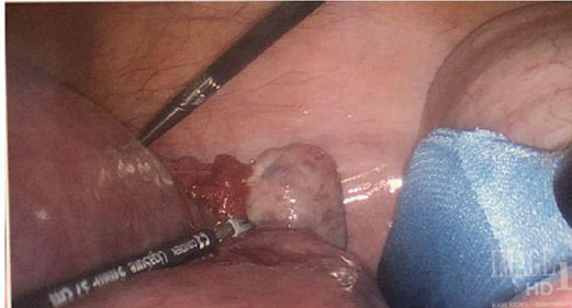
On hospital day 7, a pelvic ultrasound was obtained which showed a right ovarian mass with two echogenic nodules measuring 1.4 × 1.4 × 1.7 cm and 1 × 1 × 1.3 cm as well as peripheral calcifications. The left ovary showed no discrete mass. Of note, her first trimester ultrasound showed unremarkable adnexa. She was taken to the operating room (OR) by the gynecology team on hospital day 8, at which time she underwent an uncomplicated laparoscopic right oophorectomy. Intraoperatively, both ovaries appeared grossly normal (Figure 1, Figure 2, Figure 3).
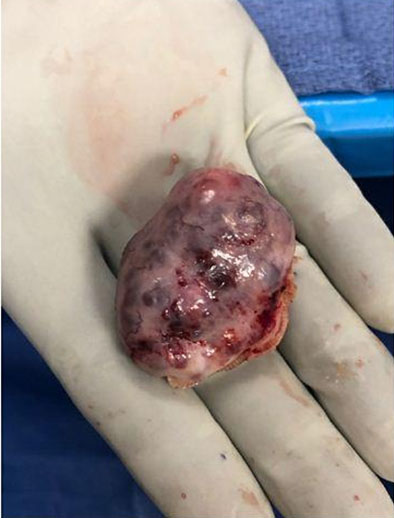
Following surgery, the working diagnosis was anti-NMDA encephalitis. Autoimmune studies ultimately returned with cerebral spinal fluid positive for NMDA antibodies at a titer of 1:32. She began additional treatment with plasma exchange (PLEX) therapy and high dose steroids. Pathology of the right ovary ultimately returned with mature neural tissue, positive for S100 and GFAP with negative anti-Neu antibodies, confirming suspicion of teratoma-associated anti-NMDA encephalitis.
After completing five plasma exchange (PLEX) treatments, her neurologic exam remained unchanged. She then received five doses of intravenous immunoglobulin (IVIg) as the next step in management. She intermittently required pressors for autonomic dysfunction and hypotension, and a tracheostomy was performed due to her prolonged intubation on hospital day 24. She continued to exhibit significant automatisms, including orofacial and hand jerking dyskinesias.
On hospital day 35, she underwent another lumbar puncture which showed improving cell counts and protein. She received a two-day course of high dose IVIg therapy and a second dose of rituximab two weeks after the initial dose, however, her neurologic status still remained unchanged. She was then restarted on PLEX therapy in hopes for improvement.
At 30 weeks and 2 days gestation, the obstetrical team was notified by nursing of concern for preterm labor. On evaluation, her cervix was 2 cm with regular contractions. Her labor was augmented by rupture of membranes and she progressed well. She ultimately had a successful delivery in the intensive care unit (ICU) with delivery of a female newborn at 30 weeks and 5 days gestation. The neonatal APGAR scores were 7 and 8. She required approximately 6 minutes of positive pressure ventilation before being weaned to continuous positive airway pressure (CPAP) and ultimately to unassisted room air on the same day as delivery.
Following preterm vaginal delivery, the patient continued to have significant automatisms. A pelvic MRI visualized a hyperstimulated left ovary and transvaginal ultrasound (US) showed an enlarging left ovarian mass, and her repeat anti-NMDA titers returned worsening at 1:128. In the setting of continued autonomic dysfunction, neurologic automatisms, enlarged left ovary, and worsening titers, gynecology was re-consulted for the possibility of ongoing production of anti-NMDA antibodies from the remaining left ovary.
After multiple discussions with the patient’s family regarding removal of the contralateral ovary and surgical menopause, her family ultimately decided to proceed with left salpingo-oophorectomy and right salpingectomy on hospital day 69; intraoperatively, the left ovary was described as “slightly enlarged with multiple small cysts that appeared hemorrhagic but were intact.” The pathology returned consistent with a second teratoma. After removal of the contralateral teratoma and initiation of intrathecal rituximab as well as cytoxan, she underwent weekly serial lumbar punctures with down-titrating titers, from 1:128 to 1:20 to 1:10 to 1:80.
Over the ten days following surgical removal of the left ovary, she was weaned off sedation and all antiepileptics except levetiracetam. She was taken off the ventilator and underwent tracheostomy collar trials. By hospital day 79, she was transferred out of the ICU to the floor.
She began to show improvement in mental status with simple commands, level of cognition, and level of alertness. Intrathecal rituximab was discontinued due to high risk of the procedure and improving mental status, but monthly Cytoxan was continued for a second dose along with high dose prednisone, levetiracetam, and phenobarbital for seizures as well as propranolol for autonomic dysfunction and tachycardia. By hospital day 99, she was able to follow commands and answer questions with single word responses; at that time, she was deemed appropriate for discharge to an acute rehabilitation facility on levetiracetam only, where she stayed for one month before returning home.

Discussion
There is a known association of anti-NMDAR encephalitis and ovarian teratoma, with the majority of affected patients being women [2]. Given the median age of onset of 23 years old, there have been several cases of severe encephalitis diagnosed during pregnancy [3]. Many of these cases describe severe neurologic impairment, including seizures, autonomic dysfunction, coma, and hypoventilation [4]. Despite severity of symptoms, the antibody mediated neurological damage is usually treatment-responsive and reversible, with many patients experiencing full recovery [4]. Treatment consists of surgical resection of the teratoma, if present, as well as immunotherapy comprised of high-dose corticosteroids, immunoglobulin, plasma exchange, and anti-inflammatory agents [2]. Case series show that anti-NMDAR encephalitis can be successfully treated during pregnancy, and that prompt surgical resection of any teratoma identified leads to a higher likelihood of complete recovery [2] as well as lower rates of relapse [4].
This case illustrates the importance of complete resection of ectopic neural tissue in recovery of an affected patient. It is estimated that approximately 60% of patients will have a tumor at time of neurologic symptoms, with ovarian teratoma being the most common tumor type [4]. In an initial case series of 98 patients, 8 had bilateral teratomas with 4 of those being synchronous, 2 having a history of previous teratoma, and 2 developing a contralateral teratoma [4]. This suggests that there is subset of patients susceptible to multiple teratoma development in the setting of anti-NDMAR encephalitis, which raises concern for potential neurologic relapse.
Ours is the first case of contralateral teratoma development occurring during the course of treatment and within a pregnancy, and demonstrates that a second teratoma can develop remarkably fast. It has been reported that the size of the teratoma does not contribute to the severity of encephalitis, making it crucial to identify even very small teratomas. In this case, it is unclear whether a miniscule teratoma existed in the contralateral ovary and went undetected by imaging and at the time of surgery, or if anti-NMDAR antibodies already in circulation triggered its rapid development. Regardless, in future cases of disease plateau or deterioration, repeat imaging and close monitoring of antibody titers should be obtained to evaluate for the possibility of a second or incompletely resected tumor. This should not be dismissed even over a short course, as in this case, development of the second teratoma was obviously visible on imaging within two months of previously normal results.
Conclusion
Removal of ovarian teratomas can be a vital component of the treatment of anti-NMDAR encephalitis. Diagnosis and treatment can be challenging with a spectrum of neurologic impacts. If the clinical course of a patient has deteriorated or plateaued, consideration should be given to recurrence or development of a new ovarian teratoma. This should be considered even over short periods of time and especially during pregnancy.
REFERENCES
1.
Dalmau J, Tüzün E, Wu HY, et al. Paraneoplastic anti-N-methyl-D-aspartate receptor encephalitis associated with ovarian teratoma. Ann Neurol 2007;61(1):25–36. [CrossRef]
[Pubmed]

2.
Motohara T, Tayama S, Narantuya D, Tashiro H, Katabuchi H. Anti-N-methyl-D-aspartate receptor encephalitis associated with ovarian teratoma: Clinical presentation, diagnosis, treatment, and surgical management. Int Canc Conf J 2013;2:121–30. [CrossRef]
3.
Joubert B, García-Serra A, Planagumà J, et al. Pregnancy outcomes in anti-NMDA receptor encephalitis: Case series. Neurol Neuroimmunol Neuroinflamm 2020;7(3):e668.
[Pubmed]
4.
Dalmau J, Gleichman AJ, Hughes EG, et al. Anti- NMDA-receptor encephalitis: Case series and analysis of the effects of antibodies. Lancet Neurol 2008;7(12):1091–8. [CrossRef]
[Pubmed]
SUPPORTING INFORMATION
Author Contributions
Catherine Boniface - Conception of the work, Design of the work, Drafting the work, Revising the work critically for important intellectual content, Final approval of the version to be published, Agree to be accountable for all aspects of the work in ensuring that questions related to the accuracy or integrity of any part of the work are appropriately investigated and resolved.
Brittany Austin - Conception of the work, Design of the work, Drafting the work, Revising the work critically for important intellectual content, Final approval of the version to be published, Agree to be accountable for all aspects of the work in ensuring that questions related to the accuracy or integrity of any part of the work are appropriately investigated and resolved.
Julio Chalela - Analysis of data, Revising the work critically for important intellectual content, Final approval of the version to be published, Agree to be accountable for all aspects of the work in ensuring that questions related to the accuracy or integrity of any part of the work are appropriately investigated and resolved.
Roger Newman - Conception of the work, Design of the work, Revising the work critically for important intellectual content, Final approval of the version to be published, Agree to be accountable for all aspects of the work in ensuring that questions related to the accuracy or integrity of any part of the work are appropriately investigated and resolved.
Guaranter of SubmissionThe corresponding author is the guarantor of submission.
Source of SupportNone
Consent StatementWritten informed consent was obtained from the patient for publication of this article.
Data AvailabilityAll relevant data are within the paper and its Supporting Information files.
Conflict of InterestAuthors declare no conflict of interest.
Copyright© 2021 Catherine Boniface et al. This article is distributed under the terms of Creative Commons Attribution License which permits unrestricted use, distribution and reproduction in any medium provided the original author(s) and original publisher are properly credited. Please see the copyright policy on the journal website for more information.


