 |
Case Report
Surgical management of a large non-obstetric vulvar hematoma: A case report
1 Department of Obstetrics and Gynecology, University Clinical Center of Kosovo, 10000 Prishtina, Kosovo
2 Faculty of Medicine, University of Prishtina “Hasan Prishtina’’, Prishtina, Kosovo
3 Department of Abdominal Surgery, University Clinical Center of Kosovo, 10000 Prishtina, Kosovo
Address correspondence to:
Vjosa A Zejnullahu
PhD, Department of Obstetrics and Gynecology, University Clinical Center of Kosovo, 10000 Prishtina,
Kosovo
Message to Corresponding Author
Article ID: 100103Z08VZ2022
Access full text article on other devices

Access PDF of article on other devices

How to cite this article
Zejnullahu VA, Zejnullahu VA, Kosumi E. Surgical management of a large non-obstetric vulvar hematoma: A case report. J Case Rep Images Obstet Gynecol 2022;8:100103Z08VZ2022.ABSTRACT
Introduction: Non-obstetric vulvar hematomas are rare emergencies and there are no guidelines with defined recommendations for the treatment. They constitute up to 0.8% of all gynecological admissions and occur as a result of perineal trauma when compression of soft vulvar tissues against the osseous planes causes damage to the vulvar vascular complex.
Case Report: We present a case of a non-obstetric vulvar hematoma in a 19-year-old woman resulting from the blunt trauma of the vulvar region.
Conclusion: Our experience suggests that surgical intervention, in the setting of an expanding vulvar hematoma, reduces length of hospital stay and minimizes associated morbidity.
Keywords: Drainage, Evacuation, Surgical approach, Trauma, Vulvar hematoma
Introduction
Vulvovaginal, paravaginal, and retroperitoneal hematomas are common in obstetric settings whereas non-obstetrics vulvar hematomas are very rare. Puerperal hematomas occur as a complication of vaginal or perineal bleeding lacerations, episiotomy, after a spontaneous injury to a blood vessels during delivery and after vaginal instrumental delivery [1].
The reported incidence of puerperal hematomas ranges from 1:300 to 1:1500 deliveries [2].
Non-obstetric vulvar hematomas occur as a result of perineal trauma when compression of soft vulvar tissues against the osseous planes causes damage to the vulvar vascular complex [3]. Therefore, vulvar trauma may result in hematomas or external bleeding. Main contributors to this complication are a rich vascular supply of the perineum and vulva as well as valveless, thin-walled female pelvis veins [4]. Numerous anastomoses with the large pelvic venous plexuses and low endurance of subcutaneous fatty tissue permit hemorrhage or massive hematomas [4].
Traumatic vulvar hematomas constitute up to 0.8% of all gynecological admissions [5] and have a reported incidence of 3.7% [6]. They may be caused by car or bicycle accidents [3], fall onto a metal object, fall from a height [7] insertion of a foreign body, sexual assault, consensual intercourse [6],[8],[9] and vulvar surgery [10].
Vulvar hematomas with or without lacerated injuries caused by snowboarding were also reported in the literature [11].
Although, trauma is the main cause of non-obstetric vulvar hematoma, one study reported vulvar hematoma secondary to spontaneous rupture of the internal iliac artery [12] and another study reported a case of vulvar hematoma with rupture of pseudoaneurysm of pudendal artery in absence of trauma [13].
Non-obstetric vulvar hematomas are common in children, pre-pubertal girls and postmenopausal women. In adult women, large fat deposits of the labia majora, protect the vulva against traumatic injury [14]. Lack of this protection in children and pre-pubertal girls is a main cause of hematoma formation after injury. Similarly, postmenopausal women because of estrogen deficiency, vulvar atrophy, and changes in skin barrier functions are more prone to hematoma formation following trauma [15].
The main symptom of vulvar hematoma is pain, described by the patient as a perineal, abdominal or buttock pain. Other symptoms include neurological, urological symptoms, difficulty walking or sitting [7],[12]. Vulvar hematomas usually present as a tender compressible mass covered by skin with purplish discoloration [16].
Initial evaluation of the patient should include prompt identification of a hematoma, assessment for vaginal, urethral, anal and bony pelvis injuries, patient resuscitation and individualized treatment [17].
We present a case of a vulvar hematoma in a 19-year-old woman resulting from the blunt trauma of the vulvar region.
Case Report
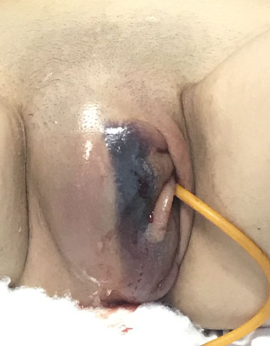
A 19-year-old woman presented to the Emergency room at the Obstetrics and Gynecology Clinic with a large vulvar hematoma following an accidental fall on a wooden object after a short episode of postural syncope. The patient was hospitalized in our Clinic due to the sudden-onset unilateral swelling, associated with severe pain and difficulty walking (Figure 1). Her personal medical history was unremarkable and she was not on any medication. She denied any history of prior sexual intercourse or sexual assault. The patient showed moderate distress on admission, because of local pain.
Clinical examination revealed a hemodynamically stable patient (blood pressure 110/80 mmHg, heart rate 80 bpm, arterial blood oxygen saturation of 99%, and an oral temperature of 37.2°C).
Genital examination revealed a tender, swollen area of approximately 12×7 cm on the entire right vulvar region. The labia was displaced to the left thus considerably deforming her genital anatomy. The patient did not present skin or mucosal wounds as well as vaginal bleeding at the moment of admission. Catherization was performed because of the urinary retention, caused by progressive labial swelling and local pain; 650 mL of clear urine was drained.
A preoperative laboratory assessment showed complete blood count levels within normal ranges (RBC 3.69 × 10⁶/mm3, hemoglobin level 11.6 g/dL, hematocrit 32.8%, WBC 10.8 × 10³/mm3, and platelet count 197×103/mm3, bleeding time 1 minute, 30 seconds, clotting time 5 minutes, 45 seconds).
Ultrasound examination of the area revealed a large blood collection in the right labia. Because of the increased pain severity despite parenterally administered pain relievers and acute expansion of the hematoma on physical examination, a decision for surgical management was made, 1 hour later (Figure 2). Prior to intervention patient consent for surgery was obtained.
Under spinal anesthesia, a vertical incision was performed over the point of maximal protrusion on the right labia, in order to evacuate hematoma. After evacuation of multiple blood clots (>500 mL) the space created by hematoma was reapproximated using five interrupted sutures (polyglactin 910) without draining or packing the hematoma cavity. No sign of further bleeding was observed (Figure 3).
Patient reported a significant decrease of the pain and distress few hours after the surgery. Patient received intravenous crystalloids, broad-spectrum antibiotic, analgesics and had an uneventful postoperative clinical course. She was discharged on the fifth postoperative day (Figure 4).
Discussion
Non-obstetric vulvar hematomas are rare and constitute up to 0.8% of all gynecological emergencies [5]. They usually occur as a result of accidents, sexual assault, and consensual coitus [6],[7],[8],[9]. Patients with vulvar hematoma in absence of the external genital trauma require prompt investigation for spontaneous vessel rupture [12],[13].
The perineal region and vulva have a rich blood supply which is clinically important, especially after birth, trauma or surgical procedures. The vulva is supplied by the branches of the internal pudendal artery including small branches to the gluteal region, inferior hemorrhoidal artery, perineal artery, and artery of the clitoris [4]. The branches of the pudendal artery are located in the superficial fascia of the anterior and posterior triangle. Therefore, propagation of bleeding is limited by Colles’ fascia in the urogenital triangle and by anal fascia in the posterior triangle [14].
In the presented case, our patient fell on an object that caused contusion of the vulvar vascular network against the pelvic bones. In this area, bleeding is restricted by Colles’ fascia and can progress to the skin because of low endurance of subcutaneous fatty tissue [14].
Arterial bleeding arises from the branches of the pudendal artery and usually results in hemodynamic instability while in a case of venous bleeding from multiple sites, identification and ligation of causative vessels are often difficult to achieve [14].
Initial assessment of the patient should include a detailed history of external trauma, genital examination, assessment for associated pelvic injuries, laboratory work-up, and diagnostic imaging. After physical examination, perineal sonography is a useful tool in the assessment of the location and expansion of the hematoma and can assist in the expectant management through serial bedside imaging [18]. For cases where retroperitoneal involvement, arteriovenous malformations or aneurysms are suspected, CT-angiography is recommended as more accurate imaging modality [14]. Guerriero et al. recommend magnetic resonance imaging (MRI) for a more detailed mapping of the lesion and exclusion of retroperitoneal involvement [19].
The literature is inconclusive regarding the best treatment approach for vulvar hematomas. However, there is a general consensus that conservative management with bed rest, pain relievers, cold compression, and bladder catheterization is recommended for small, nonexpanding vulvar hematomas [20]. These patients need close observation, to detect any vaginal or retroperitoneal involvement in a timely manner [14].
Expansion of the hematoma or hemodynamically unstable patient necessitates active surgical management, to prevent infection and necrosis caused by pressure [17]. In the presented case, surgical exploration with blood clot evacuation and primary closure was performed because of fast enlarging hematoma and severe pain. We have found this approach to be successful although drainage of the hematoma is also an alternative method.
Zahn et al, advised aggressive operative management and drainage for vulvovaginal hematomas complicating delivery [21]. Benrubi et al. recommended surgical approach when the product of the longitudinal and the transverse diameter is 15 cm or greater. Results from this study, found more subsequent operative intervention, increased complications and increased hospital stay for conservatively managed patients compared to the operatively managed group [22].
Selective angiographic embolization is a safe and effective alternative to surgical approach for pelvic hemorrhage and larger vulvar hematomas [12]. Yet, there is a lack of data comparing advantages and disadvantages of the two methods for the treatment of non-obstetric hematomas.
Conclusion
Non-obstetric vulvar hematomas are rare and they usually occur as a result of perineal trauma. Conservative management with close observation is recommended for small, nonexpanding vulvar hematomas. In the setting of a fast expanding vulvar hematomas refractory to conservative management, early surgical approach provides a rational and efficient treatment. Operative management reduces length of the hospital stay and can minimize associated morbidity.
REFERENCES
1.
Oong GC, Eruo FU. Vulvar Hematoma. 2021 Aug 14. In: StatPearls. Treasure Island (FL): StatPearls Publishing; 2022.
[Pubmed]

2.
Villella J, Garry D, Levine G, Glanz S, Figueroa R, Maulik D. Postpartum angiographic embolization for vulvovaginal hematoma. A report of two cases. J Reprod Med 2001;46(1):65–7.
[Pubmed]

3.
Virgili A, Bianchi A, Mollica G, Corazza M. Serious hematoma of the vulva from a bicycle accident. A case report. J Reprod Med 2000;45(8):662–4.
[Pubmed]

5.
Rabinerson D, Fradin Z, Zeidman A, Horowitz E. Vulvar hematoma after cunnilingus in a teenager with essential thrombocythemia: A case report. J Reprod Med 2007;52(5):458–9.
[Pubmed]

6.
Jones ISC, O’Connor A. Non-obstetric vulval trauma. Emerg Med Australas 2013;25(1):36–9. [CrossRef]
[Pubmed]

7.
Lapresa Alcalde MV, Hernández Hernández E, Bustillo Alfonso S, Doyague Sánchez MJ. Non-obstetric traumatic vulvar hematoma: Conservative or surgical approach? A case report. Case Rep Womens Health 2019;22:e00109. [CrossRef]
[Pubmed]

8.
Mangwi AA, Ebasone PV, Aroke D, Ngek LT, Nji AS. Non-obstetric vulva haematomas in a low resource setting: Two case reports. Pan Afr Med J 2019;33:314. [CrossRef]
[Pubmed]

9.
Golbasi C, Golbasi H, Bayraktar B, Ertas İE. Surgical management and clinical follow-up of post-coital severe vulvar hematoma in a 14-year-old adolescent girl: A case presentation. Acta Biomed 2021;92(S1):e2021041. [CrossRef]
[Pubmed]

10.
Theodorou G, Khomsi F, Bouzerda-Brahami K, Bouquet de Jolinière J, Feki A. Surgical management of a large postoperative vulvar haematoma following vulvar phlebectomy and ovarian vein embolization for vulvar varicose veins: A case report. Case Rep Womens Health 2020;27:e00225. [CrossRef]
[Pubmed]

11.
Kanai M, Osada R, Maruyama K, Masuzawa H, Shih HC, Konishi I. Warning from Nagano: Increase of vulvar hematoma and/or lacerated injury caused by snowboarding. J Trauma 2001;50(2):328–31. [CrossRef]
[Pubmed]

12.
Egan E, Dundee P, Lawrentschuk N. Vulvar hematoma secondary to spontaneous rupture of the internal iliac artery: Clinical review. Am J Obstet Gynecol 2009;200(1):e17–8. [CrossRef]
[Pubmed]

13.
Hong HR, Hwang KR, Kim SA, et al. A case of vulvar hematoma with rupture of pseudoaneurysm of pudendal artery. Obstet Gynecol Sci 2014;57(2):168–71. [CrossRef]
[Pubmed]

14.
Roman AS. Management of hematomas incurred as a result of obstetrical delivery. UpToDate 2021. [Available at: https://www.uptodate.com/contents/management-of-hematomas-incurred-as-a-result-of-obstetric-delivery]

15.
Farage M, Maibach H. Lifetime changes in the vulva and vagina. Arch Gynecol Obstet 2006;273(4):195–202. [CrossRef]
[Pubmed]

16.
Zahn CM, Yeomans ER. Postpartum hemorrhage: Placenta accreta, uterine inversion, and puerperal hematomas. Clin Obstet Gynecol 1990;33(3):422–31. [CrossRef]
[Pubmed]

17.
Ridgway LE. Puerperal emergency. Vaginal and vulvar hematomas. Obstet Gynecol Clin North Am 1995;22(2):275–82.
[Pubmed]

18.
Sherer DM, Stimphil R, Hellmann M, Abdelmalek E, Zinn H, Abulafia O. Transperineal sonography of a large vulvar hematoma following blunt perineal trauma. J Clin Ultrasound 2006;34(6):309–12. [CrossRef]
[Pubmed]

19.
Guerriero S, Ajossa S, Bargellini R, Amucano G, Marongiu D, Melis GB. Puerperal vulvovaginal hematoma: Sonographic findings with MRI correlation. J Clin Ultrasound 2004;32(8):415–8. [CrossRef]
[Pubmed]

20.
Propst AM, Thorp JM Jr. Traumatic vulvar hematomas: Conservative versus surgical management. South Med J 1998;91(2):144–6. [CrossRef]
[Pubmed]

21.
Zahn CM, Hankins GD, Yeomans ER. Vulvovaginal hematomas complicating delivery. Rationale for drainage of the hematoma cavity. J Reprod Med 1996;41(8):569–74.
[Pubmed]

22.
Benrubi G, Neuman CA, Nuss RC, Thompson RJ. Vulvar and vaginal hematomas: A retrospective study of conservative versus operative management. South Med J 1987;80(8):991–4. [CrossRef]
[Pubmed]

SUPPORTING INFORMATION
Acknowledgments
Authors wish to thank the patient for her consent for publication of this case report and the accompanying images.
Author ContributionsVjosa A Zejnullahu - Conception of the work, Design of the work, Acquisition of data, Analysis of data, Drafting the work, Final approval of the version to be published, Agree to be accountable for all aspects of the work in ensuring that questions related to the accuracy or integrity of any part of the work are appropriately investigated and resolved.
Valon A Zejnullahu - Revising the work critically for important intellectual content, Final approval of the version to be published, Agree to be accountable for all aspects of the work in ensuring that questions related to the accuracy or integrity of any part of the work are appropriately investigated and resolved.
Ernad Kosumi - Drafting the work, Final approval of the version to be published, Agree to be accountable for all aspects of the work in ensuring that questions related to the accuracy or integrity of any part of the work are appropriately investigated and resolved.
Guaranter of SubmissionThe corresponding author is the guarantor of submission.
Source of SupportNone
Consent StatementWritten informed consent was obtained from the patient for publication of this article.
Data AvailabilityAll relevant data are within the paper and its Supporting Information files.
Conflict of InterestAuthors declare no conflict of interest.
Copyright© 2022 Vjosa A Zejnullahu et al. This article is distributed under the terms of Creative Commons Attribution License which permits unrestricted use, distribution and reproduction in any medium provided the original author(s) and original publisher are properly credited. Please see the copyright policy on the journal website for more information.


