 |
Case Report
Primary urethral carcinoma mistaken as a caruncle: A case report
1 Department of Obstetrics and Gynecology, Kern Medical, Bakersfield, California, USA; David Geffen School of Medicine at UCLA, Los Angeles, CA, USA
2 Ross University School of Medicine, St. Michael, Barbados
3 Department of Obstetrics and Gynecology, Kern Medical, Bakersfield, California, USA
4 Department of Urology, Kern Medical, Bakersfield, California, USA
Address correspondence to:
Yufan Brandon Chen
9330 Stockdale Hwy #400, Bakersfield, CA 93311,
USA
Message to Corresponding Author
Article ID: 100115Z08YC2022
Access full text article on other devices

Access PDF of article on other devices


How to cite this article
Chen YB, Bibby S, Hirai-Adachi C, Hillyer S. Primary urethral carcinoma mistaken as a caruncle: A case report. J Case Rep Images Obstet Gynecol 2022;8:100115Z08YC2022.ABSTRACT
Introduction: Urethral caruncles are the most common benign urethral lesion in females, especially in post-menopausal women. In rare cases, primary urethral carcinoma can develop from a caruncle.
Case Report: We present a case of primary urethral carcinoma in a patient who presented with urethral tenderness and what appeared to be a benign urethral caruncle. After failing conservative therapy, she underwent excision of the lesion, which revealed moderately differentiated invasive squamous cell carcinoma. She then underwent excision of the distal urethra and anterior vaginal wall. No residual disease was discovered and she recovered well without symptoms of stress urinary incontinence.
Conclusion: Excision of a urethral caruncle is indicated in a symptomatic patient not responding to conservative therapy in order to rule out an underlying malignancy.
Keywords: Cancer, Caruncle, Dysuria, Urethra
Introduction
Urethral caruncles are the most common benign urethral lesion in females, especially in post-menopausal women. Usually found on the posterior meatus of the urethra, a caruncle can be described as a pedunculated, grossly vascular, red or violaceous lesion. Histologically, it is described as a mixed hypoplastic urothelial and squamous lining overlying a fibrotic, edematous, inflamed vascular stroma [1]. Symptoms typically include bleeding, dysuria, and a urethral mass, although obstructive urinary symptoms can occur with larger lesions. Treatment is usually conservative and includes Sitz baths, topical estrogens, anti-inflammatory medications, or steroids, though a systematic review noted there is a lack of data to guide management [2].
When symptoms are refractory to conservative therapy, excision should be considered. This report describes a rare case of primary squamous cell carcinoma arising from a urethral caruncle and clinical considerations.
Case Report
A 50-year-old primiparous post-menopausal woman presented to the clinic with complaints of dyspareunia, dysuria, and a vaginal mass for the past two years. She reported no significant past gynecologic history, including previously normal PAP tests. Pelvic examination was remarkable for a tender 3 mm urethral caruncle along the posterior urethral meatus. Vaginal atrophy was seen throughout, especially along the anterior vaginal wall and urethral meatus. At the initial visit, malignancy was deemed unlikely as the lesion did not have solid components or increased vascularity and was homogenous in appearance. Due to the patient’s discomfort, a speculum exam could not be performed and the decision was made to proceed with vaginal estrogen cream daily for two months. Upon follow-up, the patient reported no improvement in her symptoms and new onset vaginal bleeding. On examination, the vaginal atrophy had improved; however, there was no change in the urethral lesion size or tenderness. Pelvic ultrasound showed a leiomyomatous uterus with a 1.5 cm submucosal mass within the endometrial cavity. At this point, the differential diagnosis included post-menopausal bleeding from an endometrial polyp or endometrial cancer, and symptomatic urethral caruncle.
The patient underwent hysteroscopy with polypectomy of two endometrial polyps, cystourethroscopy, and excision of the urethral lesion (authors YC and CH). Cystourethroscopy confirmed a 2 mm wide base arising from the posterior portion of the distal urethra (Figure 1). Excision involved identifying the base of the lesion and placing stay stitches of 4-0 delayed absorbable suture around the base. The lesion was sharply excised flush with the urethral mucosa and the sutures were tied to achieve hemostasis. Histopathology showed benign endometrial polyps and moderately differentiated invasive squamous cell carcinoma from the urethral mass (Figure 2).
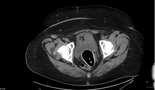
This case was reviewed at the hospital tumor board and the patient was referred to a urologist and medical oncologist for further evaluation. The patient underwent imaging with positron emission tomography-computed tomography (PET-CT), which showed no marked activity in the pelvis such as enlarged or suspicious lymph nodes. She was presented with the options of chemo-radiation therapy or distal urethrectomy and chose to undergo surgery where the distal 1 cm of the urethra and anterior vaginal wall was resected (authors YC and SH, Figure 3). Intraoperative frozen sections of the margins were negative for dysplasia or malignancy. Histopathology revealed no residual disease and therefore no additional chemotherapy or radiation treatment was recommended. The patient recovered well with no complaints of urinary incontinence. She received cancer surveillance with the plan of pelvic exams and cystourethroscopy every three months for the first year. At six months post-op there was no further evidence of disease and she remained asymptomatic.


Discussion
This case brings awareness to the possibility of malignancy arising from a urethral mass. Urethral caruncles are commonly encountered by the gynecologist; however, it is important to consider biopsy/excision when treating a symptomatic lesion that does not respond to conservative therapy. In this case, primary urethral carcinoma was diagnosed in what appeared to be a common urethral caruncle. The features of a malignant lesion include fixed mass, nodularity, ulceration, heterogeneous appearance, and large size. In the absence of these features, excision should still be considered for the relief of refractory symptoms.
Primary urethral carcinoma in a female is a rare malignancy with fewer than 100 cases diagnosed in the United States every year; however, it is an aggressive disease with 5-year survival rates of 40–60% [3]. Tumor location, particularly in the distal urethra, has been associated with improved survival (71%) compared to proximal (48%) and pan-urethral lesions (24%) [4]. Prognosis also depends on tumor stage, with 5-year survival rates of 67%, 53%, and 17% for stage II, stage III, and stage IV, respectively [5]. Malignancies occurring within a urethral lesion are often considered at least stage II because it is difficult to determine a margin from a small tissue sample.
The technical considerations for the excision of urethral caruncle includes exam under anesthesia and cystourethroscopy to visualize the stalk of the lesion. Stay sutures are placed lateral to the stalk and tied down after excision of the lesion to achieve hemostasis. A Foley catheter is left in place for 1 to 2 weeks to avoid strictures and promote wound healing.
The majority of urethral malignancies in the distal urethra are low staged and can be cured with local excision alone [6]. These tumors can be excised along with a portion of the anterior vaginal wall via a transvaginal approach [7]. One of the risks of this surgery is development of urinary incontinence if the mid-urethra or sphincter is compromised. We measure the total urethral length by marking the Foley catheter at the urethral meatus, removing the catheter, then measuring the distance from the mark to the inflated catheter balloon. The tip of a Kelly clamp is marked at 1 cm intervals, placed in the urethra, and palpated along the anterior vaginal wall to determine the margins of dissection. Using this technique, we were able to excise exactly 1.0 cm of distal urethra and maintain the patient’s continence.
There is lack of evidence to guide surveillance regimens due to the low incidence of urethral carcinoma. In patients undergoing urethra sparing surgery, we recommend routine cystourethroscopy and pelvic examinations. For those who develop urinary incontinence, conservative options such as pelvic floor physical therapy, pessaries, and urethral bulking agents should be considered. In patients who receive pelvic radiation, have significantly shortened urethra lengths, or demonstrate poor tissue quality of the anterior vaginal wall, a fascial sling or Burch urethropexy should be considered rather than a synthetic sling due to the increased risk of mesh complications.
Conclusion
Primary urethral carcinoma may present with a mass similar to a urethral caruncle. Excision of a urethral caruncle is indicated in a symptomatic patient not responding to conservative therapy in order to rule out an underlying malignancy.
REFERENCES
1.
Conces MR, Williamson SR, Montironi R, Lopez-Beltran A, Scarpelli M, Cheng L. Urethral caruncle: Clinicopathologic features of 41 cases. Hum Pathol 2012;43(9):1400–4. [CrossRef]
[Pubmed]

2.
Verma V, Pradhan A. Management of urethral caruncle – A systematic review of the current literature. Eur J Obstet Gynecol Reprod Biol 2020;248:5–8. [CrossRef]
[Pubmed]
3.
Champ CE, Hegarty SE, Shen X, et al. Prognostic factors and outcomes after definitive treatment of female urethral cancer: A population-based analysis. Urology 2012;80(2):374–81. [CrossRef]
[Pubmed]
4.
Dalbagni G, Zhang ZF, Lacombe L, Herr HW. Female urethral carcinoma: An analysis of treatment outcome and a plea for a standardized management strategy. Br J Urol 1998;82(6):835–41. [CrossRef]
[Pubmed]
5.
Derksen JW, Visser O, de la Rivière GB, Meuleman EJ, Heldeweg EA, Lagerveld BW. Primary urethral carcinoma in females: An epidemiologic study on demographical factors, histological types, tumour stage and survival. World J Urol 2013;31(1):147–53. [CrossRef]
[Pubmed]
6.
Grabstald H, Hilaris B, Henschke U, Whitmore WF Jr. Cancer of the female urethra. JAMA 1966;197(11):835–42.
[Pubmed]
7.
Anderson CB, McKiernan JM. Tumors of the urethra. In: Partin AW, Dmochowski RR, Kavoussi LR, Peters CA, Wein AJ, editors. Campbell-Walsh-Wein Urology. 12ed. Philadelphia: Elsevier; 2021. p. 1782–85.
SUPPORTING INFORMATION
Author Contributions
Yufan Brandon Chen - Conception of the work, Design of the work, Acquisition of data, Analysis of data, Drafting the work, Revising the work critically for important intellectual content, Final approval of the version to be published, Agree to be accountable for all aspects of the work in ensuring that questions related to the accuracy or integrity of any part of the work are appropriately investigated and resolved.
Sarah Bibby - Conception of the work, Design of the work, Acquisition of data, Drafting the work, Final approval of the version to be published, Agree to be accountable for all aspects of the work in ensuring that questions related to the accuracy or integrity of any part of the work are appropriately investigated and resolved.
Chihiro Hirai-Adachi - Conception of the work, Design of the work, Acquisition of data, Drafting the work, Revising the work critically for important intellectual content, Final approval of the version to be published, Agree to be accountable for all aspects of the work in ensuring that questions related to the accuracy or integrity of any part of the work are appropriately investigated and resolved.
Shahab Hillyer - Conception of the work, Design of the work, Acquisition of data, Analysis of data, Drafting the work, Revising the work critically for important intellectual content, Final approval of the version to be published, Agree to be accountable for all aspects of the work in ensuring that questions related to the accuracy or integrity of any part of the work are appropriately investigated and resolved.
Guaranter of SubmissionThe corresponding author is the guarantor of submission.
Source of SupportNone
Consent StatementWritten informed consent was obtained from the patient for publication of this article.
Data AvailabilityAll relevant data are within the paper and its Supporting Information files.
Conflict of InterestAuthors declare no conflict of interest.
Copyright© 2022 Yufan Brandon Chen et al. This article is distributed under the terms of Creative Commons Attribution License which permits unrestricted use, distribution and reproduction in any medium provided the original author(s) and original publisher are properly credited. Please see the copyright policy on the journal website for more information.


