 |
Clinical Image
Vaginal stone formation around intrauterine device in an ambulatory woman with morbid obesity
1 Department of Obstetrics and Gynecology, University of North Carolina, Chapel Hill, NC, USA
2 School of Medicine, University of North Carolina, Chapel Hill, NC, USA
Address correspondence to:
Madeline Thornton
Department of Obstetrics and Gynecology, School of Medicine, University of North Carolina, 3020 Old Clinic, CB #7570, Chapel Hill, NC 27599,
USA
Message to Corresponding Author
Article ID: 100133Z08RS2022
Access full text article on other devices

Access PDF of article on other devices


How to cite this article
Silverstein RG, Patnam R, Thornton M. Vaginal stone formation around intrauterine device in an ambulatory woman with morbid obesity. J Case Rep Images Obstet Gynecol 2022;8(2):45–48.ABSTRACT
No Abstract
Keywords: Intrauterine device, Obesity, Pelvic pain, Urinary incontinence, Vaginal stone
Case Report
A 59-year-old woman with a body mass index (BMI) of 72 kg/m2 presented to a community hospital for acute onset, severe vaginal pain. Her exam was notable for a large, textured mass in her vagina. Due to equipment weight limits, this emergency room was unable to obtain imaging and transferred her to a tertiary medical center.
Her past medical history included hypertension, atrial flutter, obstructive sleep apnea, obesity hypoventilation, type II diabetes, and chronic kidney disease. She was able to perform most of her activities of daily living, and she could ambulate with a walker but often used a wheelchair.
Her gynecologic history included stage II pelvic organ prolapse, frequent urinary tract infections (UTIs), and mixed stress, urge, and functional urinary incontinence. A specialist in female pelvic medicine and reconstructive surgery had evaluated her and advised behavioral changes as she was not felt to be a surgical candidate.
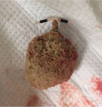
In our emergency room, we performed a physical examination on a bariatric stretcher without stirrups, requiring the assistance of five staff members. Urine covered the perineum and stretcher. We were unable to insert a speculum and palpated a 4 cm, rock hard, yellow, and brown textured mass in the distal vagina, which we then manually extracted in the emergency room. There was a copper intrauterine device (IUD) protruding from a calculus. The patient reported improvement in her symptoms and was discharged home with outpatient follow-up.
The calculus was composed of magnesium ammonium phosphate (struvite), calcium phosphate (hydroxyapatite and carbonate apatite), and magnesium phosphate trihydrate (Figure 1). It was 4.5.×4.0×3.5 cm and weighed 40 g.
The patient had a ParaGard IUD placed 20 years prior, with multiple attempts at removal, including by complex family planning specialists. Her cervix was never visualized secondary to habitus; an exam under anesthesia was deemed too dangerous.
Two years prior to presentation, a computed tomography (CT) urogram was notable for a “radiopaque device noted in the cervix, presumably related to known uterine prolapse” (Figure 2). There was no documentation on further evaluation of these findings.

Discussion
This case of vaginal stone is unique as the patient has no genitourinary abnormalities and is ambulatory. The patient’s morbid obesity was likely the primary contributing factor to the stone formation, and the stone formed around a ParaGard IUD.
The physiologic vagina is not conducive to stone formation. In order for stones to form, there typically must be: (a) significant urine; (b) outlet obstruction; and (c) an alkaline environment [1]. In this patient, vaginal urine was likely secondary to retrograde flow due to incontinence and frequent recumbent position, rather than an anatomical abnormality, which is typically seen in vaginal stones. Retrograde urinary flow is classically seen only in non-ambulatory patients [2],[3],[4]. She likely had outlet obstruction from redundant tissue rather than the usual transverse septum, imperforate hymen [5], or trauma [1]. The patient’s frequent urinary tract infections (UTIs), thought to be in part due to her habitus, could produce an alkaline environment.
Vaginal stones usually form spontaneously, but can form around vaginal mesh, retained gauze, and self-inserted items [6],[7],[8],[9],[10]. There are case reports of stones forming around IUDs such as Lippes Loops, Safe-T-Coil, and stainless-steel ring IUD, but this case is unique as there are not any reports of the IUDs currently used in the United States [11],[12],[13],[14].
This patient’s obesity likely contributed to stone formation and further affected her care. The community hospital did not have adequate imaging equipment, we did not have bariatric pelvic beds, and her IUD was unable to be removed in clinic. Additionally, providers are less likely to perform annual pelvic exams on obese patients [15].
This case highlights the clinical importance of keeping vaginal stones in the differential, following up incidental findings on imaging, and performing regular gynecologic care, even in patients for whom pelvic exams are difficult, particularly if they have specific gynecologic complaints. This case study suggests that IUD removal may be pertinent in obese patients with urinary incontinence to avoid potential complications such as vaginal stone formation, and providers should not defer IUD removal due to perceived difficulty of removal in this patient population.
Conclusion
This case demonstrates a rare complication of leaving an IUD in situ in mainly recumbent, obese women with urinary incontinence and serves as a reminder of the importance of equitable care even when challenging.
REFERENCES
1.
Avsar AF, Keskin HL, Catma T, Kaya B, Sivaslioglu A A. A large primary vaginal calculus in a woman with paraplegia. J Low Genit Tract Dis 2013;17(1):61–5. [CrossRef]
[Pubmed]

2.
Oguzkurt P, Ince E, Ezer SS, Temiz A, Demir S, Hicsonmez A. Primary vaginal calculus secondary to urethrovaginal fistula with imperforate hymen in a 6-year-old girl. J Pediatr Surg 2009;44(7):e11–3. [CrossRef]
[Pubmed]
3.
Castellan P, Nicolai M, De Francesco P, et al. Primary vaginal calculus in a woman with disability: Case report and literature review. J Endourol Case Rep 2017;3(1):182–5. [CrossRef]
[Pubmed]
4.
Lin CJ, Hsu HH, Chen CP, Wu CH, Chen HY. Huge primary vaginal stone in a recumbent woman. Taiwan J Obstet Gynecol 2005;44(1):80–2. [CrossRef]
5.
Ho TC, Lin IL. Primary vaginal stone in a young active woman. Taiwan J Obstet Gynecol 2008;47(4):457–9. [CrossRef]
[Pubmed]
6.
Malhotra N, Kumar S, Roy KK, Agarwal R, Verma V. Vaginal calculus secondary to vaginal outlet obstruction. J Clin Ultrasound 2004;32(4):204–6. [CrossRef]
[Pubmed]
7.
Dalela D, Agarwal R, Mishra VK. Giant vaginolith around an unusual foreign body—An uncommon cause of urinary incontinence in a girl. Br J Urol 1994;74(5):673–4. [CrossRef]
[Pubmed]
8.
Griffiths KM, Towers GD, Yaklic JL. Vaginal urinary calculi formation secondary to vaginal mesh exposure with urinary incontinence. Case Rep Obstet Gynecol 2017;2017:8710315. [CrossRef]
[Pubmed]
9.
Lukács T, Mohammed S. Giant secondary vaginal calculus. [Article in Hungarian]. Orv Hetil 1989;130(47):2537–38.
[Pubmed]
10.
Patankar S, Dobhada S, Bhansali M. Vesicovaginal fistula with secondary vaginal stones. J Laparoendosc Adv Surg Tech A 2006;16(4):386–9. [CrossRef]
[Pubmed]
11.
Madden A, Aslam A, Nusrat NB. A case of migrating “saf-t-coil” presenting with a vesicovaginal fistula and vesicovaginal calculus. Urol Case Rep 2016;7:17–9. [CrossRef]
[Pubmed]
12.
Beedham T, Rao K. Giant vaginal stone with embedded contraceptive device. J R Soc Med 2001;94(10):522–3. [CrossRef]
[Pubmed]
13.
Karsmakers R, Weis-Potters AE, Buijs G, Joustra EB. Chronic kidney disease after vesico-vaginal stone formation around a migrated intrauterine device. BMJ Case Rep 2010;2010:bcr1220092547. [CrossRef]
[Pubmed]
14.
Yan D, Shi Z, Wang L, Zhao X. Migration of a fractured ring IUD resulting in vesicovaginal fistula and vaginal calculus. Eur J Contracept Reprod Health Care 2018;23(5):387–9. [CrossRef]
[Pubmed]
15.
Hernandez-Boussard T, Ahmed SM, Morton JM. Obesity disparities in preventive care: Findings from the National Ambulatory Medical Care Survey, 2005–2007. Obesity (Silver Spring) 2012;20(8):1639–44. [CrossRef]
[Pubmed]
SUPPORTING INFORMATION
Acknowledgments
We would like to acknowledge and thank the patient who graciously allowed us to share her story in this article so that it may inform future gynecologic care.
Author ContributionsR Gina Silverstein - Conception of the work, Design of the work, Acquisition of data, Analysis of data, Drafting the work, Revising the work critically for important intellectual content, Final approval of the version to be published, Agree to be accountable for all aspects of the work in ensuring that questions related to the accuracy or integrity of any part of the work are appropriately investigated and resolved.
Radhika Patnam - Conception of the work, Design of the work, Acquisition of data, Analysis of data, Drafting the work, Revising the work critically for important intellectual content, Final approval of the version to be published, Agree to be accountable for all aspects of the work in ensuring that questions related to the accuracy or integrity of any part of the work are appropriately investigated and resolved.
Madeline Thornton - Conception of the work, Design of the work, Acquisition of data, Analysis of data, Drafting the work, Revising the work critically for important intellectual content, Final approval of the version to be published, Agree to be accountable for all aspects of the work in ensuring that questions related to the accuracy or integrity of any part of the work are appropriately investigated and resolved.
Guaranter of SubmissionThe corresponding author is the guarantor of submission.
Source of SupportNone
Consent StatementWritten informed consent was obtained from the patient for publication of this article.
Data AvailabilityAll relevant data are within the paper and its Supporting Information files.
Conflict of InterestAuthors declare no conflict of interest.
Copyright© 2022 R Gina Silverstein et al. This article is distributed under the terms of Creative Commons Attribution License which permits unrestricted use, distribution and reproduction in any medium provided the original author(s) and original publisher are properly credited. Please see the copyright policy on the journal website for more information.


