 |
Case Report
Vulvar adenofibroma and pregnancy: A case study, at the regional hospital of Thiès
1 Gynecology Department, Thiès Regional Hospital Center (CHART), Thiès, Senegal
2 Health Sciences Training and Research Unit of Thiès, Thiès, Senegal
3 Obstetrics and Gynecology Department, Saint-Louis Regional Hospital Center, Gaston Berger University of Saint-Louis, Senegal
4 Obstetrics and Gynecology Department, Hopital Dantec Dakar, Cheikh Anta Diop University of Dakar, Senegal
5 Obstetrics and Gynecology Department, Tivaouane Hospital Center; Health Sciences Training and Research Unit of Thiès, Senegal
Address correspondence to:
Lamine Gueye
Gynecology and Obstetrics Department of the Thiès Regional Hospital Center, 1 Avenue Malick SY Prolongée, BP: 34, Thiès,
Senegal
Message to Corresponding Author
Article ID: 100165Z08LG2023
Access full text article on other devices

Access PDF of article on other devices


How to cite this article
Gueye L, Thiam M, Thiam O, Bentefouet TL, Gassama O, Ba PA, Dabo N, Cissé ML. Vulvar adenofibroma and pregnancy: A case study, at the regional hospital of Thie?s. J Case Rep Images Obstet Gynecol 2023;9(2):51–54.ABSTRACT
Introduction: Benign vulvar epithelial tumors developing from glandular structures are rare. Given the resemblance of vulvar fibroadenomas to those of mammary locations, some authors have put forward the hypothesis of development from accessory mammary glands.
Case Report: We report a case of hyperalgesic giant vulvar fibroadenoma occurring in a 22-year-old female patient at 28 weeks, 3 days gestation. She was admitted for a painful mass on the right labia majora that had been present for two years, with a remarkable increase in volume during pregnancy. Faced with the acute vulvar pain and the discomfort caused by the mass, the decision to perform a vulvar lumpectomy was made and carried out semi-urgently, leading to the excision of a mass weighing 1103 grams. Microscopic examination revealed an adenofibroma. The patient gave birth vaginally at 38 weeks of gestation to a male baby weighing 3115 grams, 10 weeks after lumpectomy.
Conclusion: Fibroadenoma is a benign tumor located in most cases in the breast, its location in the vulva is rare and can lead to a diagnostic delay. Surgical treatment by complete excision allows healing.
Keywords: Pregnancy, Senegal, Thiès, Vulvar fibroadenoma
Introduction
Benign vulvar epithelial tumors developing from glandular structures are rare [1]. Given the resemblance of vulvar fibroadenomas to those of mammary location, some authors [2],[3] have put forward the hypothesis of development from accessory mammary glands based on topographical arguments and due to the presence of glands resembling mammary acini in the periphery. We report a case of hyperalgesic giant vulvar fibroadenoma occurring in a 22-year-old female patient carrying a pregnancy of 28 weeks, 3 days of gestation. The objective was to describe the clinical, pathological characteristics and prognosis of this association.
Case Report
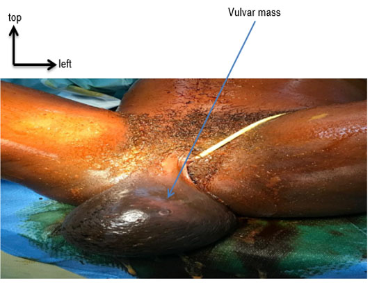
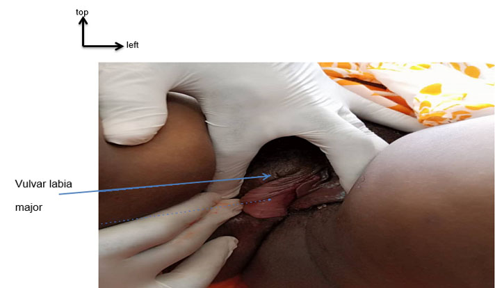
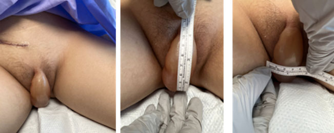
This was a 22-year-old female patient, primigravida, with no significant medical or surgical history, admitted for a painful mass on the right labia majora that had been present for two years, with an increase in volume during pregnancy. The examination revealed a mass on the right major lip, measuring 17 cm × 15 cm. This mass was firm in consistency, painful on mobilization with an orange peel appearance and a wide pedicle (Figure 1). There was no ulceration or inguinal lymphadenopathy. The obstetric examination revealed a uterine height of 27 cm, regular fetal heart sounds, and normal breasts without palpable masses. On vaginal examination the cervix was posterior, long, softened, closed, presentation was cephalic and not engaged.
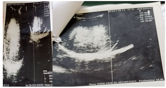
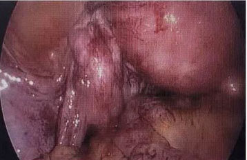
The ultrasound showed a viable, single intrauterine pregnancy of 28 weeks and 3 days of gestation associated with a vulvar mass estimated at 1166 cc of tissue appearance without significant vascularization (Figure 2). A biopsy could not be performed. We highlighted the need for a biopsy first; however, only we would have to wait 8 weeks before obtaining the definitive anatomopathological results, and given the disabling nature and the pain caused by the mass, we decided after evaluating the avascular nature of the mass to perform surgery 3 days after hospitalization.
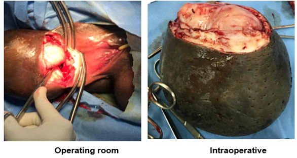
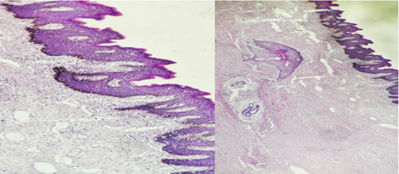
The decision to perform a vulvar lumpectomy was made (Figure 3) semi-urgently, allowing the excision of a mass weighing 1103 grams (Figure 4). The anatomopathological study showed on macroscopy of the specimen weighing 1103 grams and measuring 14×14×9. When cut, the slices showed a well-defined, non-encapsulated nodular formation measuring 12 cm long axis with a firm, elastic consistency, grayish white in color, and a fleshy appearance.
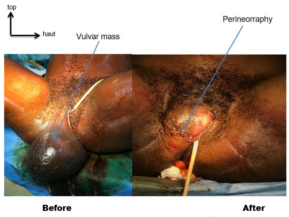
This appearance was suggestive of a fibroadenoma of the vulva (Figure 5). The evolution was favorable without recurrence (Figure 6). The patient gave birth vaginally at 38 weeks of gestation to a male baby weighing 3115 grams, 10 weeks after lumpectomy. A preventive episiotomy contralateral to the scar was performed during delivery.
Discussion
Adenofibromas located in the vulva are extremely rare [1]. Their origin remains debated and despite their macroscopic characteristics of a benign lesion, a histological study with relevant immunohistochemical techniques must be carried out to have a precise diagnosis and to rule out other vulvar tumors. The association of giant vulvar adenofibroma and pregnancy is exceptional. For this case we report, the patient was young, aged 22 years. Romero [1] published an unusual case of vulvar fibroadenoma in a 29-year-old woman who had presented for two years with a vulvar mass causing dyspareunia and post-coital bleeding.
The particularity in our context was the late diagnosis with limited means of exploration. While magnetic resonance imaging (MRI) can provide the best soft tissue resolution, ultrasound can also be used in the assessment to some extent [4]. It allowed us to appreciate the characteristics of the mass and the insignificant vascularization of the pedicle. Semi-emergency surgery during pregnancy due to the disabling nature of the vulvar mass was performed. The decision to perform a biopsy before excision of a vulvar mass should also be considered. A biopsy would be less invasive, although there may be sampling and interpretation errors [5]. It would delay timely management in our work context.
This surgery is easy because these tumors are always well limited, not encapsulated, pedunculated, and easily enucleated [3],[6]. Some are solid [3] but most are cystic and have a papillary architecture, differentiating them from vulvar fibroadenomas.
The route of delivery was independent of the vulvar lumpectomy scar. In our case, the patient went into labor spontaneously at 38 weeks of gestation and gave birth vaginally to a child of normal weight.
Conclusion
Fibroadenoma is a benign tumor located in most cases in the breast, its location in the vulva is rare and can lead to a diagnostic delay. The association of vulvar fibroadenoma and pregnancy is an exceptional situation. Pathology plays an important role in the positive diagnosis. Surgical treatment by complete excision allows healing.
REFERENCES
1.
Romero Rojas N. Vulvar fibroadenoma: Report of a case. Rev Peru Ginecol Obstet 2022;68(3). [CrossRef]

2.
Siegler AM, Gordon R. Fibroadenoma in a supernumerary breast of the vulva. Am J Obstet Gynecol 1951;62(6):1367–9. [CrossRef]
[Pubmed]
3.
Foushee JH, Pruitt AB Jr. Vulvar fibroadenoma from aberrant breast tissue. Report of 2 cases. Obstet Gynecol 1967;29(6):819–23.
[Pubmed]
4.
Hernandez Lopez AL, Manandhar S, Dubow L. Pregnancy-unrelated fibroadenoma in ectopic breast tissue in the axilla and vulva: A case report. Case Rep Womens Health 2020;28:e00255. [CrossRef]
[Pubmed]
5.
Cohen Sacher B. The normal vulva, vulvar examination, and evaluation tools. Clin Obstet Gynecol 2015;58(3):442–52. [CrossRef]
[Pubmed]
6.
Assor D, Davis JB. Multiple apocrine fibroadenomas of the anal skin. Am J Clin Pathol 1977;68(3):397–9. [CrossRef]
[Pubmed]
SUPPORTING INFORMATION
Author Contributions
Lamine Gueye - Conception of the work, Design of the work, Acquisition of data, Analysis of data, Revising the work critically for important intellectual content, Final approval of the version to be published, Agree to be accountable for all aspects of the work in ensuring that questions related to the accuracy or integrity of any part of the work are appropriately investigated and resolved.
M Thiam - Conception of the work, Design of the work, Revising the work critically for important intellectual content, Final approval of the version to be published, Agree to be accountable for all aspects of the work in ensuring that questions related to the accuracy or integrity of any part of the work are appropriately investigated and resolved.
O Thiam - Conception of the work, Design of the work, Drafting the work, Final approval of the version to be published, Agree to be accountable for all aspects of the work in ensuring that questions related to the accuracy or integrity of any part of the work are appropriately investigated and resolved.
TL Bentefouet - Conception of the work, Design of the work, Acquisition of data, Analysis of data, Drafting the work, Revising the work critically for important intellectual content, Final approval of the version to be published, Agree to be accountable for all aspects of the work in ensuring that questions related to the accuracy or integrity of any part of the work are appropriately investigated and resolved.
O Gassama - Conception of the work, Design of the work, Acquisition of data, Analysis of data, Drafting the work, Revising the work critically for important intellectual content, Final approval of the version to be published, Agree to be accountable for all aspects of the work in ensuring that questions related to the accuracy or integrity of any part of the work are appropriately investigated and resolved.
PA Ba - Conception of the work, Design of the work, Acquisition of data, Analysis of data, Drafting the work, Revising the work critically for important intellectual content, Final approval of the version to be published, Agree to be accountable for all aspects of the work in ensuring that questions related to the accuracy or integrity of any part of the work are appropriately investigated and resolved.
N Dabo - Conception of the work, Design of the work, Acquisition of data, Analysis of data, Drafting the work, Revising the work critically for important intellectual content, Final approval of the version to be published, Agree to be accountable for all aspects of the work in ensuring that questions related to the accuracy or integrity of any part of the work are appropriately investigated and resolved.
ML Cissé - Conception of the work, Design of the work, Acquisition of data, Analysis of data, Drafting the work, Revising the work critically for important intellectual content, Final approval of the version to be published, Agree to be accountable for all aspects of the work in ensuring that questions related to the accuracy or integrity of any part of the work are appropriately investigated and resolved.
Guaranter of SubmissionThe corresponding author is the guarantor of submission.
Source of SupportNone
Consent StatementWritten informed consent was obtained from the patient for publication of this article.
Data AvailabilityAll relevant data are within the paper and its Supporting Information files.
Conflict of InterestAuthors declare no conflict of interest.
Copyright© 2023 Lamine Gueye et al. This article is distributed under the terms of Creative Commons Attribution License which permits unrestricted use, distribution and reproduction in any medium provided the original author(s) and original publisher are properly credited. Please see the copyright policy on the journal website for more information.
