 |
Case Report
Phenotypic variant of diffuse uterine leiomyomatosis presenting as cellular leiomyoma with retroperitoneal extension: A case report
1 Assistant Professor, Department of Obstetrics and Gynaecology, Sri Madhusudan Sai Institute of Medical Sciences and Research, Muddenahalli, Chikkaballapur, Karnataka, India
2 Associate Professor, Department of Pathology, Sri Madhusudan Sai Institute of Medical Sciences and Research, Muddenahalli, Chikkaballapur, Karnataka, India
Address correspondence to:
Akkamahadevi C Hiremath
Assistant Professor, Department of Obstetrics and Gynaecology, Sri Madhusudan Sai Institute of Medical Sciences and Research, Muddenahalli, Chikkaballapur 562101, Karnataka,
India
Message to Corresponding Author
Article ID: 100232Z08AH2026
Access full text article on other devices

Access PDF of article on other devices


How to cite this article
Hiremath AC, Muniswamy B, Gururaj BC. Phenotypic variant of diffuse uterine leiomyomatosis presenting as cellular leiomyoma with retroperitoneal extension: A case report. J Case Rep Images Obstet Gynecol 2026;12(1):54–58.ABSTRACT
Introduction: Diffuse uterine leiomyomatosis (DUL) is a rare benign smooth-muscle disorder characterized by diffuse myometrial involvement and uterine enlargement. Its radiologic appearance may mimic malignant or extra-uterine disease, creating diagnostic uncertainty. We report an unusual case of DUL with retroperitoneal extension presenting as a giant abdomino-pelvic mass.
Case Report: A nulliparous woman in her 40s presented with abdominal pain and dyspepsia for six months with recent weight loss, without menstrual complaints. Examination revealed a markedly distended abdomen with a large abdomino-pelvic mass. Serum CA-125 was elevated (276 U/mL), while other tumor markers were within normal limits. Magnetic resonance imaging demonstrated a 26 × 16.5 × 17.5 cm T2-isointense mass occupying the abdomen and pelvis with extra-peritoneal/retroperitoneal insinuation. Differential diagnoses included diffuse leiomyomatosis, leiomyosarcoma, and ovarian neoplasm. Treatment options were discussed, and exploratory laparotomy, was performed with multidisciplinary team. Intraoperatively, a large fleshy mass with multiple projections arising from a normal uterus was identified, extending into the retroperitoneal and paravesical spaces. The appearance differed from diffuse uterine leiomyomatosis, with no evidence of peritoneal leiomyomatosis. She underwent total abdominal hysterectomy with bilateral salpingo-oophorectomy. Postoperatively she developed sudden hypotension requiring emergency re-exploration during which around 1500 mL blood clots were evacuated and a small bleeder of 2 mm in retroperitoneal space was identified and secured. Histopathology revealed a spindle cell neoplasm with increased cellularity, while immunohistochemistry showed Ki-67 <5% and negative S100 excluding malignancy. At one year follow-up, the patient was asymptomatic with no evidence of recurrence.
Conclusion: Diffuse uterine leiomyomatosis is a rare benign entity that may present as a massive abdomino-pelvic mass with atypical retroperitoneal extension, closely mimicking malignant disease. Preoperative diagnosis remains challenging despite advanced imaging. Histopathology and immunohistochemistry are essential for definitive diagnosis. Multidisciplinary surgical management and careful postoperative monitoring are crucial for favorable outcomes.
Keywords: Diffuse uterine leiomyomatosis, Immunohistochemistry, Phenotypic variant, Retroperitoneum
Introduction
Diffuse uterine leiomyomatosis is a benign condition with multiple poorly circumscribed smooth-muscle nodules occupying the myometrium in different proportions. Cellular leiomyoma is a variant of uterine leiomyoma presenting with increased smooth muscle cellularity with benign cytological features and architecture.
We report an unusual phenotypic variant of DUL spectrum disease manifesting as a large multilobulated cellular leiomyoma with retroperitoneal extension and minimal menstrual symptoms, posing significant diagnostic and intraoperative challenges. This case report was prepared in accordance with the CARE guidelines.
Case Report
A nulliparous woman in her 40s presented with 6 months of insidious, dull-aching abdominal pain and three months of dyspepsia. She reported no menstrual complaints.
Over two months, she noted reduced appetite and a 6-kg weight loss. On examination, she appeared cachectic [body mass index (BMI) 18 kg/m2]. The abdomen was markedly distended by a term sized uterine mass arising from the pelvis (Figure 1). There was no clinical ascites.
Per-vaginal examination revealed the cervix pulled up behind the pubic symphysis with fullness in the posterior fornix. The uterus could not be felt separately. Per-rectal examination showed a firm anterior rectal wall bulge with free mucosa.
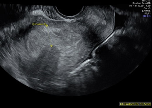
Laboratory parameters were normal except CA-125 Elevated at 276 U/mL. CA 19.9, Alpha Feto Protein (AFP), Lactate Dehydrogenase (LDH), and Carcinoembryonic Antigen (CEA) were within normal limits. Abdomino-pelvic magnetic resonance imaging (MRI) demonstrated a large ill-defined T2-isointense abdomino-pelvic mass measuring 26 × 16.5 × 17.5 cm, extending into extraperitoneal spaces. The cervix was not separately visualized. The lesion abutted major vessels posteriorly without clear invasion (Figure 2A and Figure 2B). No ascites or significant lymphadenopathy was noted. Given cachexia, rapid abdominal enlargement, and elevated CA-125, ovarian malignancy was considered. However, absence of ascites and lymphadenopathy lowered suspicion of advanced ovarian cancer. Imaging favored smooth-muscle pathology, but leiomyosarcoma could not be excluded. After counselling, exploratory laparotomy was planned with multidisciplinary backup.
Surgical Management and Postoperative Course
Through a midline vertical incision, a large multilobulated fleshy mass with multiple projections was identified. The lesion arose from an otherwise normal-appearing uterus (Figure 3) and extended into retroperitoneal and paravesical spaces. No peritoneal implants were seen. Frozen section analysis suggested a benign lesion. Total abdominal hysterectomy with bilateral salpingo-oophorectomy was performed. Lymphadenectomy was not undertaken. A clear plane of cleavage was achieved in retroperitoneal areas. The right ureter was embedded within the mass and carefully dissected free. The specimen weighed 3.4 kg. One unit packed red blood cells was transfused intraoperatively.
Six hours postoperatively, she developed tachycardia and abdominal distension. Rapid deterioration prompted re-exploration. Approximately 1500 mL of clots were evacuated from the intra and retroperitoneal space. A 2-mm spurting retroperitoneal arterial bleeder was identified and secured. She received 4 units packed red blood cells, 4 units fresh frozen plasma, and 4 units cryoprecipitate. After intensive monitoring and supportive care, she recovered and was discharged on postoperative day 10.
Histopathology and Immunohistochemistry
On histopathological examination, microscopically, the tumor comprised oval elongated to spindle looking cells with bland nuclear features and tapered cytoplasmic ends. These cells were arranged in interlacing fascicles and in whorl pattern. At foci the tumor cells separated by edema and proteinaceous fluid, resulting in a trabecular, nested architecture.
Mitoses 2–3 per 10 HPF. Necrosis was absent. Spindle cell neoplasm with differentials including cellular leiomyoma and other malignant spindle cell neoplasms like malignant peripheral nerve sheath tumor (retroperitoneal origin) were considered and immunohistochemistry with Smooth Muscle Actin (SMA), S100 and Ki-67 proliferative index were done. Smooth Muscle Actin was diffusely positive in the tumor cells confirming the smooth muscle origin of the lesion favoring cellular leiomyoma. Ki-67 index was less than 5% ruling out malignancy, and S100 was negative ruling out neural origin in a retroperitoneal mass (Figure 4A, Figure 4B, Figure 4C). These findings supported a diagnosis of cellular leiomyoma within the spectrum of diffuse uterine leiomyomatosis.
Follow-up at six months and 1 year showed no recurrence clinically or on ultrasonography.


Discussion
Diffuse uterine leiomyomatosis is an uncommon benign smooth-muscle disorder characterized by diffuse myometrial involvement. Classical presentation includes heavy menstrual bleeding and infertility. Our patient’s lack of menstrual symptoms and presence of constitutional symptoms made diagnosis challenging. The gross intraoperative appearance was unusual. Instead of diffuse uterine enlargement alone, a dominant multilobulated fleshy mass with extensive retroperitoneal extension was present, while the uterus appeared externally normal. This suggests a phenotypic variant within the DUL spectrum. Magnetic resonance imaging (MRI) is an important imaging tool in evaluation of soft tissue tumors of abdomen and pelvis and is preferred over computed tomography (CT) scan as it provides superior soft tissue contrast, identifies tissue composition and differentiates benign from malignant lesions. There may be alteration in signal characteristics when degenerative changes and cellular variants are noted [1],[2]. Intraoperative findings of, retroperitoneal extension and poor cervical delineation lead to suspicion of malignancy. Fibroids are well-defined, low T2 signal masses with homogeneous enhancement whereas irregular or ill-defined margins with heterogeneous T2 signals, high T1 signal for hemorrhage, restricted diffusion on DWI may raise suspicion of leiomyosarcoma [3]. Conclusive diagnosis is obtained only after histopathology and immunohistochemistry examination.
Management of DUL is individualized. Fertility-preserving approaches include modified myomectomy techniques and uterine artery occlusion strategies [4],[5],[6]. However, in extensive disease with retroperitoneal involvement, conservative surgery may be technically hazardous and hemorrhagic. In this patient, hysterectomy was appropriate given age, lack of fertility desire, disease extent, and intraoperative complexity. The postoperative hemorrhage underscores a key surgical lesson. Retroperitoneal dissection carries risk of concealed bleeding. Even after apparent hemostasis, small arterial bleeders may lead to significant hemoperitoneum. Vigilant monitoring, prompt recognition, and immediate re-exploration are lifesaving. Histopathological and immunohistochemistry examination for confirmation of diagnosis was critical to rule out malignancy and for follow-up.
Learning Points:
- Diagnostic overlap between benign and malignant smooth-muscle tumors.
- Importance of multidisciplinary surgical planning.
- Need for blood product preparedness.
- Role of histopathology in definitive diagnosis.
- Vigilance for concealed retroperitoneal bleeding.
- Postoperative hemorrhage in retroperitoneal dissections may present abruptly.
Conclusion
Our case highlights the challenges in diagnosis of a phenotypic variant of DUL. Among the three leiomyomatous conditions of uterus—cellular leiomyoma, diffuse uterine leiomyomatosis, and diffuse peritoneal leiomyomatosis, cellular leiomyoma can mimic sarcoma due to increased cellularity. Magnetic resonance imaging supported by histopathological and immunohistochemistry can facilitate the accurate diagnosis and aid in the customized management and follow up of atypical DUL spectrum immunohistochemistry can facilitate the accurate diagnosis and aid in the customized management and follow up of atypical DUL spectrum.
REFERENCES
1.
Silva CA, Rosa F, Rito M, Cunha TM. Diffuse leiomyomatosis: A rare cause of a diffusely enlarged uterus. Radiol Case Rep 2022;17(5):1536–9. [CrossRef]
[Pubmed]

2.
Nougaret S, Cunha TM, Benadla N, Neron M, Robbins JB. Benign uterine disease: The added role of imaging. Obstet Gynecol Clin North Am 2021;48(1):193–214. [CrossRef]
[Pubmed]
3.
Ren HM, Wang QZ, Wang JN, Hong GJ, Zhou S, Zhu JY, et al. Diffuse uterine leiomyomatosis: A case report and review of literature. World J Clin Cases 2022;10(24):8797–804. [CrossRef]
[Pubmed]
4.
Kweon S, Park J, Sim Y, Kwack JY, Kwon YS. Clinical outcomes of conservative surgery for diffuse uterine leiomyomatosis: Preliminary experience of 17 cases in a single center. J Clin Med 2023;12(24):7638. [CrossRef]
[Pubmed]
5.
Dai YX, Feng FZ, Leng JH, Shi HH, Cheng NH, Wan XR, et al. Imaging features and clinical analysis of diffuse uterine leiomyomatosis cases. [Article in Chinese]. Zhonghua Yi Xue Za Zhi 2020;100(29):2263–7. [CrossRef]
[Pubmed]
6.
Zhang Z, Yang H, Pan R. Revolutionizing diffuse uterine leiomyomatosis treatment: A case report and literature review on “no-distension” hysteroscopic myomectomy with thoracic tissue forceps. Int J Gynaecol Obstet 2025;168(1):87–93. [CrossRef]
[Pubmed]
SUPPORTING INFORMATION
Acknowledgments
The authors would like to thank the Departments of Radiology, Pathology, and Anesthesiology for their valuable support in the diagnosis and management of this case. They also acknowledge the contribution of the operating room staff and multidisciplinary surgical team involved in patient care.
Author ContributionsAkkamahadevi C Hiremath - Conception of the work, Design of the work, Acquisition of data, Analysis of data, Drafting the work, Revising the work critically for important intellectual content, Final approval of the version to be published, Agree to be accountable for all aspects of the work in ensuring that questions related to the accuracy or integrity of any part of the work are appropriately investigated and resolved.
Brunda Muniswamy - Acquisition of data, Revising the work critically for important intellectual content, Final approval of the version to be published, Agree to be accountable for all aspects of the work in ensuring that questions related to the accuracy or integrity of any part of the work are appropriately investigated and resolved.
Vivek Tubagere Gururaj - Acquisition of data, Analysis of data, Revising the work critically for important intellectual content, Final approval of the version to be published, Agree to be accountable for all aspects of the work in ensuring that questions related to the accuracy or integrity of any part of the work are appropriately investigated and resolved.
Guaranter of SubmissionThe corresponding author is the guarantor of submission.
Source of SupportNone
Consent StatementWritten informed consent was obtained from the patient for publication of this article.
Data AvailabilityAll relevant data are within the paper and its Supporting Information files.
Conflict of InterestAuthors declare no conflict of interest.
Copyright© 2026 Akkamahadevi C Hiremath et al. This article is distributed under the terms of Creative Commons Attribution License which permits unrestricted use, distribution and reproduction in any medium provided the original author(s) and original publisher are properly credited. Please see the copyright policy on the journal website for more information.


