
 |
Case Report
Laparoscopic-assisted uterine evacuation of 14-week pregnancy complicated by obstructing cervical malignancy
1 Department of Obstetrics, Gynecology, and Reproductive Sciences, Division of Complex Family Planning, University of Pittsburgh Medical Center, Pittsburgh, PA, United States
2 Department of Obstetrics, Gynecology, and Reproductive Sciences, Division of Minimally Invasive Gynecologic Surgery, University of Pittsburgh Medical Center, Pittsburgh, PA, United States
3 Department of Obstetrics, Gynecology, and Reproductive Sciences, Division of Maternal-Fetal Medicine, University of Pittsburgh Medical Center, Pittsburgh, PA, United States
4 Department of Obstetrics, Gynecology, and Reproductive Sciences, University of Pittsburgh Medical Center, Pittsburgh, PA, United States
Address correspondence to:
Monica L Kao
MD, MPH, Department of Obstetrics, Gynecology, and Reproductive Sciences, UPMC Magee-Womens Hospital, 300 Halket St, Suite 2333, Pittsburgh, PA 15222,
United States
Message to Corresponding Author
Article ID: 100235Z08MK2026
Access full text article on other devices

Access PDF of article on other devices


How to cite this article
Kao ML, Zeccola AM, Emery SP, Guido R, Uhm S, Chen BA. Laparoscopic-assisted uterine evacuation of 14-week pregnancy complicated by obstructing cervical malignancy. J Case Rep Images Obstet Gynecol 2026;12(2):5–9.ABSTRACT
Introduction: Treatment of cervical cancer in pregnant patients presents uniquely complex challenges. Treatment considerations include—but are not limited to—disease stage, histological subtype, prognostic implications of delaying treatment, gestational age, other obstetric comorbid conditions, and a patient’s personal priorities and values around continuation versus termination of pregnancy.
Case Report: We present a unique case of laparoscopic-assisted pregnancy termination at 14 weeks for a patient with newly diagnosed stage IIIC1 cervical cancer to expedite cancer treatment. Her friable, obstructing cervical mass precluded the usual approach of cervical dilation and subsequent uterine evacuation or medical termination of pregnancy. This case report describes multidisciplinary, collaborative efforts to integrate each of these factors into a cohesive treatment plan for a patient with locally advanced cervical cancer diagnosed in the second trimester. A minimally invasive approach to uterine evacuation allowed for quicker initiation of cancer treatment while reducing surgical risk and facilitating a shorter recovery period.
Conclusion: We present a complex case of a patient with a new obstructing cervical cancer in the second trimester. Multidisciplinary team management and creative problem solving allowed us to perform a laparoscopic, ultrasound-guided uterine evacuation to terminate the pregnancy allowing for expedited cancer treatment.
Keywords: Abortion, Cervical cancer, Laparoscopic, Minimally invasive approach, Uterine evacuation
Introduction
Cervical cancer is the most common gynecological cancer diagnosed in pregnancy, with an incidence of 1.5–12 cases per 100,000 pregnancies [1],[2]. Treatment of cervical cancer in pregnant patients presents uniquely complex challenges. Considerations include disease stage, histological subtype, prognostic implications of delaying treatment, gestational age, other comorbid conditions, and the patient’s personal priorities and values around continuation versus termination of pregnancy. This case report describes multidisciplinary efforts to integrate these factors into a cohesive treatment plan for a patient with locally advanced cervical cancer in the second trimester. A minimally invasive approach to uterine evacuation shortened the delay to initiation of cancer treatment while reducing surgical risk and facilitating a shorter recovery period.
Case Report
Case
A 33-year-old woman (Gravida 3, Para 2) presented for evaluation of first-trimester bleeding nine days after a positive home pregnancy test and an unsure last menstrual period. Her medical history included two uncomplicated term vaginal deliveries, anemia, and prior tobacco use. She denied a history of abnormal Pap tests, with a reportedly normal result five years prior. She described bright red bleeding and passage of small clots with intermittently heavier bleeding during intervals of greater activity, in which she would saturate up to two pads per hour. She also reported copious watery vaginal discharge.
In the office during her initial presentation for care, a speculum examination demonstrated pooling of yellow-brown watery discharge admixed with blood. A large fungating mass measuring approximately 7 cm in diameter with an irregular verrucose surface filled the apex of the vagina and bled easily upon contact with sloughing of cervical tissue. This tissue was sent as a biopsy to pathology and was histologically reported to be moderately to poorly differentiated invasive squamous cell carcinoma. Parametrial involvement was suspected based on her exam.
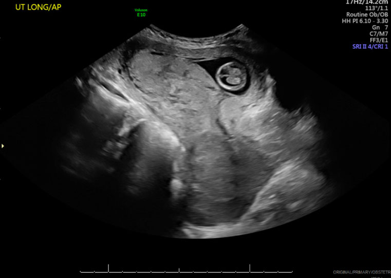
By ultrasonography, the pregnancy was estimated to be 12 weeks and 6 days gestation. An irregularly shaped, heterogenous, and vascular mass measuring 101 × 62 × 86 mm was seen within the cervix (Figure 1).
The patient was seen quickly thereafter for consultation with Gynecologic Oncology and Radiation Oncology. Imaging options to evaluate extent of local disease [magnetic resonance imaging (MRI) with contrast] and to assess for metastatic disease [positron emission tomography (PET) and computed tomography (CT)] were limited in the setting of ongoing pregnancy. The patient was counseled on pregnancy options and implications for oncologic therapy and disease prognosis. Treatment of locally advanced cervical cancer without evidence of distant metastasis typically involves concomitant chemotherapy with external beam radiation therapy and high dose-rate (HDR) intracavitary brachytherapy (chemoRT); however, this option is not compatible with pregnancy continuation. Pregnancy continuation with neoadjuvant chemotherapy followed by definitive radiation after delivery was an option, though typically reserved for earlier stage disease as delaying oncologic therapy in favor of pregnancy continuation would be life-limiting. Following careful consideration, the patient and her family opted to pursue pregnancy termination to expedite initiation of chemoradiotherapy.
Decision-making and surgical planning
We convened a multidisciplinary team including Gynecologic Oncology, Radiation Oncology, Maternal-Fetal Medicine, Complex Family Planning, and Minimally Invasive Gynecologic Surgery. Medication abortion with mifepristone/misoprostol was not an option, given this patient’s presentation in the second trimester and risk of potentially catastrophic bleeding from the cervical malignancy. The friable, obstructing cervical mass also precluded the usual approach to procedural abortion with cervical dilation and uterine evacuation. We considered feticidal injection with intracardiac potassium chloride (KCl) to interrupt further growth of the pregnancy, followed by initiation of chemotherapy and radiation to reduce tumor burden to where dilation and evacuation (D&E) could be performed. However, given the size of the cervical mass, it was unlikely that this would achieve adequate tumor regression and restoration of normal anatomy to allow for a transcervical approach to uterine evacuation. Thus, uterine evacuation via hysterotomy was planned.
We then considered timing of uterine evacuation relative to external beam therapy. Given the risk of hematologic side effects with chemoRT, risks and benefits of uterine evacuation prior to chemoRT were weighed against those of evacuation after chemoRT completion. The anticipated length of daily chemoRT was 25 fractions over a period of five weeks, followed by a brachytherapy boost. Delaying evacuation until after completion of chemoRT with prolonged retention of the demised fetus carried increased risks for spontaneous labor, bleeding, coagulopathy, and infection in an immunosuppressed patient [3]. Spontaneous abortion would require emergent laparotomy and hysterotomy since a transcervical approach was not feasible. An open abdominal incision within the irradiated field would require radiation therapy to be delayed for several weeks to allow for adequate wound healing. Interruption of external beam therapy would have a more detrimental impact upon its curative potential and patient prognosis than delayed initiation of chemoRT, thus early uterine evacuation prior to chemoRT was favored.
Given the importance of minimizing treatment delays, we proposed a laparoscopic approach to hysterotomy and uterine evacuation via suction curettage. Compared with laparotomy, laparoscopic incisions carried lesser risks of wound breakdown and poor wound healing. A minimally invasive approach would then allow for a shorter delay to initiation of radiation therapy. This technique was felt to be technically feasible, as a laparoscopic approach to hysterotomy previously had been described in the literature (although this was criticized as being a poor substitute for dilation and evacuation when a transcervical approach is possible) [4],[5]. If this approach were to be unsuccessful and require conversion to laparotomy, the plan would default to initiating chemoRT after two weeks as previously discussed.
Clinical course
At 14 weeks 0 days gestation, we performed ultrasound-guided fetal intracardiac KCl injection to interrupt pregnancy progression, reduce uterine blood flow, and facilitate uterine evacuation planned for five days later. Subsequent PET-CT showed a large, Fluorodeoxyglucose (FDG) avid mass measuring 8.7 × 9.7 × 9.1 cm extending into the vagina with no definite regional metastatic disease. Based on exam and PET-CT, she was thought to have at least Stage IIB cervical cancer, but final staging would be determined based on post-procedure MRI. The patient was prophylactically transfused with three units of packed red blood cells for a preoperative hemoglobin of 6.8 g/dL. Interventional Radiology performed preoperative gelfoam uterine artery embolization to reduce bleeding risk given the large cervical tumor and potential uterine hypervascularity.
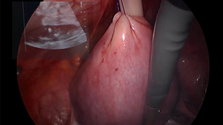
Complex Family Planning and Minimally Invasive Gynecologic Surgery specialists performed the surgery. Ports were placed strategically to optimize access to the uterus and limit the number of incisions within the radiation field. We utilized a 5 mm port in the left upper quadrant for the laparoscope, a 10 mm supraumbilical port for the laparoscopic ultrasound probe, a 10 mm infraumbilical port for the suction cannula, and a 5 mm left lower quadrant assistant port. After injection of a dilute vasopressin solution, we placed a figure-of-eight stitch of 2-0 polydioxanone suture (PDS) inferior to the planned hysterotomy site and brought it through the 10 mm infraumbilical port to provide countertraction. We used monopolar hook cautery to make a 1 cm vertical hysterotomy and carried it down to the level of the fetal membranes. We removed the 10 mm infraumbilical port and replaced it with a 12 mm flexible suction cannula. We placed traction on the stay suture to elevate the uterus toward the anterior abdominal wall and provide countertraction as the suction cannula was advanced through the hysterotomy. Using the laparoscopic ultrasound probe on the posterior uterus, we evacuated the uterus with suction curettage under direct ultrasound guidance (Figure 2). Complete evacuation of uterine contents was confirmed by laparoscopic and transabdominal ultrasound and gross inspection of the products of conception. The patient’s postoperative course was uncomplicated and she initiated chemoradiotherapy as planned on postoperative day #5. Pelvic MRI on post-operative day #10 demonstrated the large heterogeneous mass measuring 7.7 × 6.7 × 4.7 cm with bilateral parametrial extension and invasion into the upper 1/3 of the posterior vagina. The right internal iliac lymph nodes were enlarged and she was noted to have a necrotic left external inguinal lymph node. This was consistent with a diagnosis of Stage IIIC1 cervical cancer.
She completed chemoradiation with an initially encouraging treatment response (Figure 3). She underwent planned HDR brachytherapy with plans for 3-month post-treatment imaging and follow-up with Radiation Oncology and Gynecologic Oncology.

Discussion
Given the relatively low incidence of cervical cancer diagnosed in pregnancy, there is a paucity of evidence to guide standardization of treatment for pregnant patients with cervical cancer [3],[6],[7],[8]. Current recommendations for treatment of locally advanced cervical cancer discourage any treatment delays when diagnosis occurs during the first or second trimesters [9],[10]. Pregnancy may impose limitations on available treatment options resulting in considerable prognostic implications. This case underscores the importance of protecting access to pregnancy termination as a crucial treatment consideration in oncologic care. It also highlights multidisciplinary collaboration in the management of medically and surgically complex patients. Through collaborative efforts between Gynecologic Oncology, Radiation Oncology, Maternal-Fetal Medicine, Complex Family Planning, and Minimally Invasive Gynecologic Surgery, we were able to envision, construct, and execute this innovative and novel management plan.
In patients for whom a laparotomy would result in delayed treatment, increased risk for postoperative complications, and increased morbidity, a less invasive approach should be considered [3],[9]. Laparoscopic hysterotomy has been described in case reports as a less invasive alternative to laparotomy in the setting of failed induction termination and in resection of cesarean scar ectopic pregnancy; however, we describe a novel technique using suction curettage to evacuate the uterus through a small hysterotomy when transcervical approach is not feasible [4],[5],[11]. The safety of dilation and evacuation has been well established, and whenever possible, a D&E by a trained surgeon with the requisite skillset is safer and far less invasive than a hysterotomy performed via laparotomy or laparoscopy [5],[6],[7],[8],[9],[10],[11],[12]. However, when structural or anatomic considerations preclude the usual approach with cervical dilation, a laparoscopic approach to uterine evacuation with intra-abdominal ultrasound can be considered a safe and minimally invasive alternative to hysterotomy via laparotomy.
Conclusion
In this patient with locally advanced cervical cancer, a minimally invasive approach for pregnancy termination was associated with lower surgical risk, faster recovery time, and shorter delay to treatment initiation with external beam radiation therapy and radiosensitizing chemotherapy.
REFERENCES
1.
Al-Halal H, Kezouh A, Abenhaim HA. Incidence and obstetrical outcomes of cervical intraepithelial neoplasia and cervical cancer in pregnancy: A population-based study on 8.8 million births. Arch Gynecol Obstet 2013;287(2):245–50. [CrossRef]
[Pubmed]

2.
Creasman WT. Cancer and pregnancy. Ann N Y Acad Sci 2001;943:281–6. [CrossRef]
[Pubmed]
3.
Hunter MI, Tewari K, Monk BJ. Cervical neoplasia in pregnancy. Part 2: Current treatment of invasive disease. Am J Obstet Gynecol 2008;199(1):10–8. [CrossRef]
[Pubmed]
4.
Baekelandt J, Bosteels J. Laparoscopic hysterotomy for a failed termination of pregnancy: A first case report with demonstration of a new surgical technique. J Minim Invasive Gynecol 2015;22(4):544. [CrossRef]
[Pubmed]
5.
Creinin MD, Nejad BM. Laparoscopic hysterectomy for failed labor induction abortion is neither frugal nor innovative. J Minim Invasive Gynecol 2015;22(5):918. [CrossRef]
[Pubmed]
6.
Han SN, Mhallem Gziri M, Van Calsteren K, Amant F. Cervical cancer in pregnant women: Treat, wait or interrupt? Assessment of current clinical guidelines, innovations and controversies. Ther Adv Med Oncol 2013;5(4):211–9. [CrossRef]
[Pubmed]
7.
Morice P, Narducci F, Mathevet P, Marret H, Darai E, Querleu D, et al. French recommendations on the management of invasive cervical cancer during pregnancy. Int J Gynecol Cancer 2009;19(9):1638–41. [CrossRef]
[Pubmed]
8.
Morice P, Uzan C, Gouy S, Verschraegen C, Haie-Meder C. Gynaecological cancers in pregnancy. Lancet 2012;379(9815):558–69. [CrossRef]
[Pubmed]
9.
Korenaga TRK, Tewari KS. Gynecologic cancer in pregnancy. Gynecol Oncol 2020;157(3):799–809. [CrossRef]
[Pubmed]
10.
Sood AK, Sorosky JI, Mayr N, Krogman S, Anderson B, Buller RE, et al. Radiotherapeutic management of cervical carcinoma that complicates pregnancy. Cancer 1997;80(6):1073–8. [CrossRef]
[Pubmed]
11.
Pickett CM, Minalt N, Higgins OM, Bernard C, Kasper KM. A laparoscopic approach to cesarean scar ectopic pregnancy. Am J Obstet Gynecol 2022;226(3):417–9. [CrossRef]
[Pubmed]
12.
Grimes DA, Schulz KF. Morbidity and mortality from second-trimester abortions. J Reprod Med 1985;30(7):505–14.
[Pubmed]
SUPPORTING INFORMATION
Acknowledgments
Paniti Sukumvanich, MD
Parul N Barry, MD
Francesca L Facco, MD
Jamie Lesnock, MD
Kathryn Thomas, MD
Monica L Kao - Acquisition of data, Drafting the work, Revising the work critically for important intellectual content, Final approval of the version to be published, Agree to be accountable for all aspects of the work in ensuring that questions related to the accuracy or integrity of any part of the work are appropriately investigated and resolved.
Alison M Zeccola - Acquisition of data, Drafting the work, Revising the work critically for important intellectual content, Final approval of the version to be published, Agree to be accountable for all aspects of the work in ensuring that questions related to the accuracy or integrity of any part of the work are appropriately investigated and resolved.
Stephen P Emery - Revising the work critically for important intellectual content, Final approval of the version to be published, Agree to be accountable for all aspects of the work in ensuring that questions related to the accuracy or integrity of any part of the work are appropriately investigated and resolved.
Richard Guido - Revising the work critically for important intellectual content, Final approval of the version to be published, Agree to be accountable for all aspects of the work in ensuring that questions related to the accuracy or integrity of any part of the work are appropriately investigated and resolved.
Suji Uhm - Revising the work critically for important intellectual content, Final approval of the version to be published, Agree to be accountable for all aspects of the work in ensuring that questions related to the accuracy or integrity of any part of the work are appropriately investigated and resolved.
Beatrice A Chen - Revising the work critically for important intellectual content, Final approval of the version to be published, Agree to be accountable for all aspects of the work in ensuring that questions related to the accuracy or integrity of any part of the work are appropriately investigated and resolved.
Guaranter of SubmissionThe corresponding author is the guarantor of submission.
Source of SupportNone
Consent StatementWritten informed consent was obtained from the patient for publication of this article.
Data AvailabilityAll relevant data are within the paper and its Supporting Information files.
Conflict of InterestAuthors declare no conflict of interest.
Copyright© 2026 Monica L Kao et al. This article is distributed under the terms of Creative Commons Attribution License which permits unrestricted use, distribution and reproduction in any medium provided the original author(s) and original publisher are properly credited. Please see the copyright policy on the journal website for more information.


