|
Clinical Image
Twin-to-twin transfusion syndrome: One placenta, two colors
1 Resident in Obstetrics and Gynecology, Department of Obstetrics and Gynecology, Hospital de Faro, Local Health Unit of Algarve, Faro, Portugal
2 Consultant in Obstetrics and Gynecology, Department of Obstetrics and Gynecology, Hospital de Faro, Local Health Unit of Algarve, Faro, Portugal
3 Graduated Consultant in Gynecology and Obstetrics, Department of Gynecology and Obstetrics, Hospital de Faro, Unidade Local de Saúde do Algarve, Faro, Portugal
Address correspondence to:
Dinis Correia Mateus
Department of Obstetrics and Gynecology, Hospital de Faro, Local Health Unit of Algarve, Rua Leão Penedo, 8000 Faro,
Portugal
Message to Corresponding Author
Article ID: 100237Z08DM2026
Access full text article on other devices

Access PDF of article on other devices


How to cite this article
Mateus D, Edral A, Ferreira Â. Twin-to-twin transfusion syndrome: One placenta, two colors. J Case Rep Images Obstet Gynecol 2026;12(2):15–18.ABSTRACT
No Abstract
Keywords: Antenatal surveillance, Fetal demise, Monochorionic diamniotic pregnancy, Twin-to-twin transfusion syndrome
Case Report
A 39-year-old Caucasian woman, gravida 7 para 5, with a history of untreated psoriasis, trigeminal neuralgia, and significant tobacco use (20 cigarettes/day), presented at 18+1 weeks of gestation. Laboratory evaluation demonstrated gestational diabetes, and ultrasound revealed a monochorionic diamniotic (MCDA) twin pregnancy with normal amniotic fluid volumes, both fetuses in breech presentation with visible bladders, and an estimated fetal weight discordance of 6%, middle cerebral artery Doppler findings were within normal limits. The patient did not attend the scheduled ultrasound examination at 20 weeks’ gestation.
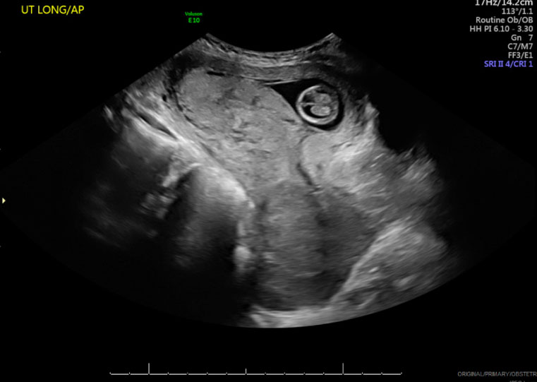
At 22 weeks, on a routine appointment, ultrasound revealed intrauterine fetal demise of both twins. One fetus exhibited subcutaneous edema with a visible bladder, while the co-twin was adherent to the uterine wall in anhydramnios with no visible bladder, consistent with Quintero stage V.
Labor was medically induced, and vaginal delivery was uneventful. Two female fetuses were expelled: the first, pale, weighing 270 g, and the second, plethoric, weighing 405 g (severe intertwin fetal weight discordance: 33.3%).
The fetopathological examination verified a MCDA twin gestation exhibiting morphological characteristics consistent with the clinical diagnosis of twin-to-twin transfusion syndrome (TTTS). Both fetuses were female; one displayed plethoric features with marked vascular congestion and edema (Twin 1: Recipient), whereas the other presented with diffuse pallor (Twin 2: Donor). The internal organs of the recipient showed an approximate 33% increase in weight compared to the donor, with an even more pronounced discrepancy in cardiac mass. No structural malformations or tissue dysplasias were identified in either fetus. The placenta exhibited chorionic villi with accelerated maturation, along with areas of pronounced congestion and vascular ectasia. No histopathological evidence of inflammatory or infectious processes was observed. The cariotype was normal for both fetus (46, XX) and infectious serologies were all negative.
Figure 1 depicts a pathognomonic image of TTTS, emphasizing the critical role of careful monitoring in MCDA pregnancies for timely detection and management of such complications.

Discussion
Monozygotic twins account for approximately 30% of spontaneous twin pregnancies, with nearly two-thirds being monochorionic (MC). In MCDA gestations, vascular anastomoses between the fetal circulations are almost universally present, establishing the basis for complications such as TTTS and twin anemia-polycythemia sequence (TAPS) [1],[2]. Twin-to-twin transfusion syndrome is a severe complication unique to MC twin pregnancies, resulting from unbalanced blood flow through placental vascular anastomoses. It affects approximately 10–15% of MCDA pregnancies [3], typically in the mid-trimester, and remains associated with high perinatal morbidity and mortality, particularly when diagnosis and intervention are delayed [4]. The present case illustrates an advanced and rapidly progressive form of TTTS culminating in intrauterine fetal demise of both twins at 22 weeks’ gestation (Figure 1). The condition is characterized by discordant amniotic fluid volumes and fetal growth, with the donor twin showing hypovolemia and uteroplacental insufficiency, and the recipient twin exhibiting hypervolemia and cardiac dysfunction [5], has seen in this case report.
At the 18-week ultrasound examination, the pregnancy showed no clear diagnostic criteria for TTTS, with normal amniotic fluid volumes, visible bladders in both fetuses, normal middle cerebral artery Doppler findings, and only mild fetal weight discordance (6%). However, MC pregnancies are characterized by dynamic hemodynamic changes, and TTTS may develop rapidly over a short period. While TTTS may develop at any point during pregnancy, diagnosis is most frequently established in the second trimester [6]. The absence of the scheduled 20-week ultrasound likely contributed to the delayed diagnosis, highlighting the importance of strict surveillance protocols in MC pregnancies, with ultrasound assessment recommended every two weeks from 16 weeks onward.
The ultrasound findings at 22 weeks were highly suggestive of advanced TTTS, corresponding to Quintero stage V, characterized by fetal demise. The marked discordance in fetal appearance after delivery, with one pale and growth-restricted fetus and the co-twin plethoric and heavier, reflects the chronic imbalance in fetoplacental transfusion. The donor twin typically develops hypovolemia, oliguria, oligohydramnios, anemia, and growth restriction, while the recipient twin may develop hypervolemia, polycythemia, cardiac overload, hydrops fetalis, and ultimately cardiac failure. In this case, the presence of subcutaneous edema in one fetus suggests hydrops, a finding associated with severe cardiovascular decompensation [3],[4].
The severe fetal weight discordance observed after delivery (33.3%) further supports the chronic and progressive nature of the transfusion imbalance. Although early ultrasound findings were relatively reassuring, the subsequent rapid deterioration emphasizes that TTTS can evolve unpredictably, even in pregnancies without initial alarming features. This reinforces the need for patient counseling regarding adherence to follow-up and the potentially rapid progression of MC twin complications.
Currently, fetoscopic laser photocoagulation of placental vascular anastomoses is the standard treatment for Quintero stage II–IV TTTS diagnosed before 26 weeks of gestation, as it directly addresses the underlying placental vascular imbalance and significantly improves perinatal survival and neurological outcomes compared with serial amnioreduction or expectant management [4],[7]. Nevertheless, alternative therapeutic options may be considered in selected clinical scenarios. Serial amnioreduction may be performed when fetoscopic laser surgery is unavailable, technically unfeasible, or contraindicated, although it does not correct the underlying pathophysiology and is associated with inferior outcomes. Septostomy, which aims to equalize amniotic fluid volumes by creating a communication between the amniotic sacs, has largely fallen out of favor because of the increased risk of cord entanglement and the lack of evidence demonstrating superiority over laser therapy [7]. Selective cord occlusion may be indicated when one fetus has a lethal anomaly or a very poor prognosis that threatens the survival of the co-twin. Similarly, selective fetal reduction (fetocide) may be considered in highly selected cases involving severe fetal compromise or major structural or chromosomal abnormalities. The choice of treatment should be individualized according to gestational age, Quintero stage, fetal condition, local expertise, and parental preferences, and should ideally be undertaken in specialized fetal therapy centers [7]. Unfortunately, in the present case, the diagnosis was established only after fetal demise, precluding therapeutic intervention.
Regardless of the treatment performed, pregnancies complicated by TTTS require intensive antenatal surveillance in specialized fetal medicine centers. According to the International Society of Ultrasound in Obstetrics and Gynecology (ISUOG) Practice Guidelines, all uncomplicated MCDA pregnancies should undergo ultrasound surveillance every two weeks from 16 weeks' gestation until delivery to facilitate early detection of TTTS and other MC complications [7]. Following the diagnosis of TTTS and particularly after fetoscopic laser photocoagulation, follow-up should be individualized according to disease severity and treatment response, with ultrasound examinations generally performed weekly during the early post-procedural period. Surveillance should include assessment of fetal growth, amniotic fluid volumes, fetal bladder filling, and Doppler studies of the umbilical artery, middle cerebral artery, and ductus venosus, with fetal echocardiography performed when clinically indicated, particularly in the recipient twin [7]. Maternal follow-up is equally important to detect obstetric complications, monitor post-procedural recovery, and provide appropriate counselling and psychological support. Close multidisciplinary surveillance is essential to optimize perinatal outcomes, detect recurrent or subsequent complications such as twin anemia-polycythemia sequence (TAPS) or selective fetal growth restriction, and determine the optimal timing of delivery.
This case also highlights the importance of strict adherence to the recommended surveillance schedule for MCDA pregnancies, as delayed diagnosis may preclude fetal intervention and result in devastating perinatal outcomes.
Conclusion
This case highlights the aggressive nature of TTTS and reinforces the importance of strict adherence to the recommended surveillance schedule for MC pregnancies. Early diagnosis is crucial, as delayed recognition may preclude fetal therapy and lead to catastrophic perinatal outcomes. Prompt referral to specialized fetal medicine centers is essential to enable timely intervention and optimize maternal and fetal outcomes.
REFERENCES
1.
Society for Maternal-Fetal Medicine (SMFM); Miller RS, Miller JL, Monson MA, Porter TF, Obi?an SG, et al. Society for Maternal-Fetal Medicine Consult Series #72: Twin-twin transfusion syndrome and twin anemia-polycythemia sequence. Am J Obstet Gynecol 2024;231(4):B16–37. [CrossRef]
[Pubmed]

2.
Durbin SA. A Sonographer’s perspective: Quintero staging system for twin-to-twin transfusion syndrome in monochorionic twins. Journal of Diagnostic Medical Sonography 2011;27(3):122–5. [CrossRef]
3.
WAPM Consensus Group on Twin-to-Twin Transfusion; Baschat A, Chmait RH, Deprest J, Gratacós E, Hecher K, et al. Twin-to-twin transfusion syndrome (TTTS). J Perinat Med 2011;39(2):107–12. [CrossRef]
[Pubmed]
4.
Miller JL. Twin to twin transfusion syndrome. Transl Pediatr 2021;10(5):1518–29. [CrossRef]
[Pubmed]
5.
Fisk NM, Galea P. Twin-twin transfusion-as good as it gets? N Engl J Med 2004;351(2):182–4. [CrossRef]
[Pubmed]
6.
Society for Maternal-Fetal Medicine; Simpson LL. Twin-twin transfusion syndrome. Am J Obstet Gynecol 2013;208(1):3–18. [CrossRef]
[Pubmed]
7.
Khalil A, Sotiriadis A, Baschat A, Bhide A, Gratacós E, Hecher K, et al. ISUOG Practice Guidelines (updated): Role of ultrasound in twin pregnancy. Ultrasound Obstet Gynecol 2025;65(2):253–76. [CrossRef]
[Pubmed]
SUPPORTING INFORMATION
Acknowledgments
The authors would like to thank Fernanda Vilela for her valuable collaboration in the collection of the clinical images used in this manuscript. The authors acknowledge the use of ChatGPT by OpenAI for English language review and editing of the discussion section of this manuscript.
Author ContributionsDinis Correia Mateus - Conception of the work, Design of the work, Acquisition of data, Analysis of data, Drafting the work, Revising the work critically for important intellectual content, Final approval of the version to be published, Agree to be accountable for all aspects of the work in ensuring that questions related to the accuracy or integrity of any part of the work are appropriately investigated and resolved.
Ana Edral - Conception of the work, Design of the work, Acquisition of data, Analysis of data, Drafting the work, Revising the work critically for important intellectual content, Final approval of the version to be published, Agree to be accountable for all aspects of the work in ensuring that questions related to the accuracy or integrity of any part of the work are appropriately investigated and resolved.
Ângela Ferreira - Conception of the work, Design of the work, Acquisition of data, Analysis of data, Drafting the work, Revising the work critically for important intellectual content, Final approval of the version to be published, Agree to be accountable for all aspects of the work in ensuring that questions related to the accuracy or integrity of any part of the work are appropriately investigated and resolved.
Guaranter of SubmissionThe corresponding author is the guarantor of submission.
Source of SupportNone
Consent StatementWritten informed consent was obtained from the patient for publication of this article.
Data AvailabilityAll relevant data are within the paper and its Supporting Information files.
Conflict of InterestAuthors declare no conflict of interest.
Copyright© 2026 Dinis Mateus et al. This article is distributed under the terms of Creative Commons Attribution License which permits unrestricted use, distribution and reproduction in any medium provided the original author(s) and original publisher are properly credited. Please see the copyright policy on the journal website for more information.