
 |
Case Report
Uterine rupture after gunshot wound with protrusion of intact amniotic sac and fetal long bones
1 Medical Student, University of Florida College of Medicine, Gainesville, FL, USA
2 MD, Clinical Assistant Professor, Department of Obstetrics and Gynecology, University of Florida College of Medicine, Gainesville, FL, USA
Address correspondence to:
Michelle Larzelere
MD, 1600 SW Archer Road, Gainesville, FL 32608,
USA
Message to Corresponding Author
Article ID: 100239Z08SZ2026
Access full text article on other devices

Access PDF of article on other devices


How to cite this article
Zettler S, Larzelere M. Uterine rupture after gunshot wound with protrusion of intact amniotic sac and fetal long bones. J Case Rep Images Obstet Gynecol 2026;12(2):25–27.ABSTRACT
Introduction: Intrapartum gunshot wounds (GSWs) to the abdomen or pelvis can cause significant injury and mortality to pregnant persons and their fetuses. Rapid imaging and emergency laparotomy are often necessary for diagnosis and treatment.
Case Report: A 14-year-old G0P0 patient at 18 weeks gestation presented status post GSW with imaging concerning for a ballistic-induced uterine rupture with fetal demise and hemoperitoneum. At the time of exploratory laparotomy, 500 cc of hemoperitoneum was noted with fetal long bones penetrating through a 5 cm rupture in the uterine wall. The amniotic sac remained intact. An en caul delivery of the fetus was performed as well as uterine curettage. Ballistic injury was noted to the left Fallopian tube and omentum. No other intra-abdominal injuries were noted. Postoperatively, the patient experienced no surgical complications and was discharged on postoperative day 10.
Conclusion: This case highlights a unique complication of an intrapartum GSW leading to uterine rupture and fetal demise at 18 weeks.
Keywords: Hemoperitoneum, Intrapartum gunshot wound, Pregnancy, Uterine trauma
Introduction
Trauma is the leading cause of nonobstetric related maternal death [1]. Uterine rupture secondary to trauma affects 0.6% of obstetric traumas with a fetal mortality rate that approaches 100% [2]. Rapid imaging and emergency laparotomy are often necessary for diagnosis and treatment. Here we present a case of uterine rupture and fetal demise secondary to a gunshot wound (GSW).
Case Report
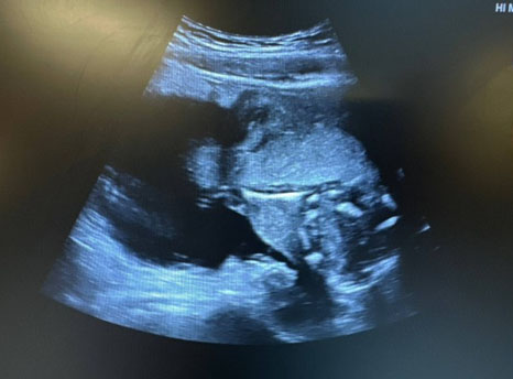
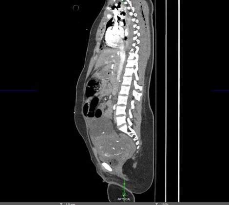
A 14-year-old G0P0 patient at 18 weeks gestation presented to the emergency department with gunshot injuries of unknown caliber and range to the right chest, back, arm, ribs 4–6, and left pelvis. At the hospital, the patient exhibited unstable vital signs [pulse: 89 BPM, blood pressure (BP) 150/107 mmHg, SpO2: 68%] and was therefore sedated and intubated. She had a chest tube placed with 1 L of bloody output and subsequently received 2 units of fresh frozen plasma and 2 units of packed red blood cells. A bedside pelvic ultrasound confirmed an intrauterine fetal demise with fetal long bones that appeared to protrude through the uterine wall (Figure 1 and Figure 2). The amniotic sac was noted to be intact. Additionally, a hypoechoic area adjacent to this rupture and hypotension suggested a collection of blood within the abdomen. These findings were confirmed on computed tomography (CT) scan (Figure 3 and Figure 4). There were no foreign bodies within the abdomen or pelvis seen on the CT to suggest retained bullets. Given these findings, an emergency exploratory laparotomy was performed jointly with both obstetric and trauma surgery teams present
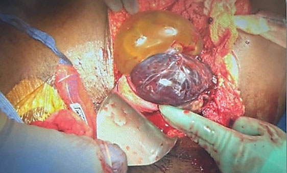
Upon entry via a midline vertical incision, a 5 cm uterine rupture from the uterine fundus to the left cornua was noted. The amniotic sac was intact and protruding through the defect (Figure 1). Roughly 500 mL of hemoperitoneum was removed with no active bleeding noted. Surgeons found two penetrating wounds overlying the left psoas muscle with intact iliac vessels and ureters. There was no solid organ, intestinal, mesenteric, or diaphragmatic injury observed, hemodynamics remained stable, and psoas bleeding was controlled with direct pressure. An en caul delivery of the fetus and placenta was performed (Figure 5). Banjo curettage was used to remove any remaining pregnancy tissue. The uterus was then closed in three layers, an initial layer of 0-Vicryl followed by an imbricating layer of 0-Vicryl and finally a baseball stitch of 3-0 Vicryl. Following the closure of the uterus, a left salpingectomy was performed as it was transected at the level of the cornua. She had no immediate postoperative complications, was transferred to the pediatric intensive care unit (PICU) in stable condition, and was discharged home in stable condition on postoperative day 10.


Discussion
Gunshot wounds during pregnancy are not uncommon in the United States—roughly 7% of pregnancies will be complicated by a traumatic injury, 4% of which come from GSWs [1]. Gunshot wounds to the abdomen of a pregnant individual are especially dangerous, causing 70% of fetuses to sustain an injury and up to 65% of pregnancies leading to fetal demise [3]. This case highlights an unusual presentation of fetal demise from a GSW, as uterine rupture occurred with preservation of an uninjured amniotic sac and fetus. The trauma and uterine rupture led to demise of the fetus.
Conclusion
This is a rare case of uterine trauma from a GSW resulting in the protrusion of fetal long bones from the uterus within the amniotic sac and fetal demise. In this case, it is likely that the ballistic led to the uterine rupture without injuring the amniotic sac or fetus.
REFERENCES
1.
Shipchandler FT, Huntley ES, Holder TF, Ali T, Behnia F, Chauhan SP, et al. Maternal and neonatal outcomes of gunshot wounds in pregnancy: A systematic review of case reports. Am Surg 2024;90(2):279–91. [CrossRef]
[Pubmed]

2.
Pearlman MD, Tintinalli JE, Lorenz RP. Blunt trauma during pregnancy. N Engl J Med 1990;323(23):1609–13. [CrossRef]
[Pubmed]
3.
Asaad M, Acuff C, Shoukralla M, Takala H, Arynova B, Chang G. A rare case of traumatic gunshot wound to fetal cranium at 25 weeks gestation: A case report. Trauma Case Rep 2024;53:101092. [CrossRef]
[Pubmed]
SUPPORTING INFORMATION
Author Contributions
Savannah Zettler - Conception of the work, Design of the work, Acquisition of data, Analysis of data, Drafting the work, Revising the work critically for important intellectual content, Final approval of the version to be published, Agree to be accountable for all aspects of the work in ensuring that questions related to the accuracy or integrity of any part of the work are appropriately investigated and resolved.
Michelle Larzelere - Conception of the work, Design of the work, Acquisition of data, Analysis of data, Drafting the work, Revising the work critically for important intellectual content, Final approval of the version to be published, Agree to be accountable for all aspects of the work in ensuring that questions related to the accuracy or integrity of any part of the work are appropriately investigated and resolved.
Guaranter of SubmissionThe corresponding author is the guarantor of submission.
Source of SupportNone
Consent StatementWritten consent was unable to be obtained from the patient but identifying information was not included in this report.
Data AvailabilityAll relevant data are within the paper and its Supporting Information files.
Conflict of InterestAuthors declare no conflict of interest.
Copyright© 2026 Savannah Zettler et al. This article is distributed under the terms of Creative Commons Attribution License which permits unrestricted use, distribution and reproduction in any medium provided the original author(s) and original publisher are properly credited. Please see the copyright policy on the journal website for more information.


