|
Case Report
A large androgen-producing ovarian mass presenting with progressive virilization in a young woman
1 Department of Gynecology and Obstetrics, Unidade Local de Saúde do Alto Minho, Viana do Castelo, Portugal
2 Department of Gynecology and Obstetrics, Instituto Português de Oncologia do Porto, Porto, Portugal
Address correspondence to:
Maria Rui Torres
Unidade Local de Saúde do Alto Minho, Rua Dr. Elisiário Monteiro Nº81, Póvoa de Varzim,
Portugal
Message to Corresponding Author
Article ID: 100240Z08MT2026
Access full text article on other devices

Access PDF of article on other devices


How to cite this article
Torres MR, Gomes MS, Pires MC. A large androgen-producing ovarian mass presenting with progressive virilization in a young woman. J Case Rep Images Obstet Gynecol 2026;12(2):28–31.ABSTRACT
Introduction: Sertoli–Leydig cell tumors (SLCT) are rare ovarian sex cord–stromal neoplasms that may produce androgens and cause progressive virilization. Recognition of this clinical presentation is important for establishing an early diagnosis and guiding appropriate treatment in young women.
Case Report: A 23-year-old nulligravid woman presented with progressive abdominal enlargement and marked hirsutism over 12 months. Her menstrual cycles were regular, and she denied other symptoms suggestive of hyperandrogenism, including acne and alopecia. Physical examination revealed marked virilization and a large abdominopelvic mass. Imaging demonstrated a predominantly solid right ovarian mass measuring approximately 20 × 15 × 10 cm. Total and free testosterone were markedly elevated at 517 and 1.94 ng/dL, respectively, while DHEA-S, 17-hydroxyprogesterone and cortisol were within normal limits; other tumor markers, including CA-125, were also unremarkable. The patient underwent right adnexectomy with omentectomy and peritoneal cytological analysis by laparotomy. Histopathological examination confirmed a moderately differentiated SLCT, International Federation of Gynecology and Obstetrics (FIGO) stage IA, with no lymphovascular invasion and negative peritoneal cytology. Molecular analysis identified two pathogenic/probably pathogenic somatic DICER1 variants. The patient remained under clinical surveillance for more than three years, with no evidence of disease recurrence.
Conclusion: Sertoli–Leydig cell tumors should be considered in young women presenting with progressive virilization and an ovarian mass. Early recognition of androgen excess may facilitate diagnosis and allow fertility-sparing treatment in selected patients with early-stage disease.
Keywords: Hirsutism, Ovarian neoplasm, Sertoli–Leydig cell tumor, Virilization
Introduction
Abdominopelvic masses in young women encompass a wide differential diagnosis, ranging from benign functional cysts to malignant ovarian neoplasms. Although most ovarian tumors in this age group are benign, the presence of endocrine manifestations such as virilization should raise suspicion for hormonally active tumors. Sertoli–Leydig cell tumors account for less than 0.5% of all ovarian neoplasms and are characterized by androgen secretion, often leading to rapid-onset hirsutism, voice deepening, and clitoromegaly [1].
Case Report
A 23-year-old nulligravid woman with no relevant medical or surgical history presented to the emergency department with progressive abdominal enlargement and marked hirsutism over 12 months. Her menstrual cycles were regular, and she denied other symptoms suggestive of hyperandrogenism, including acne and alopecia.
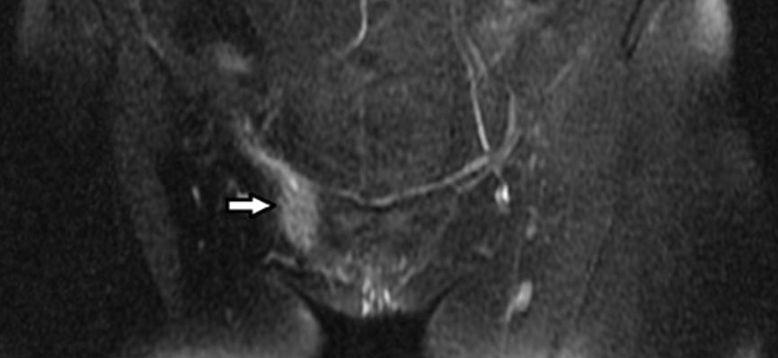
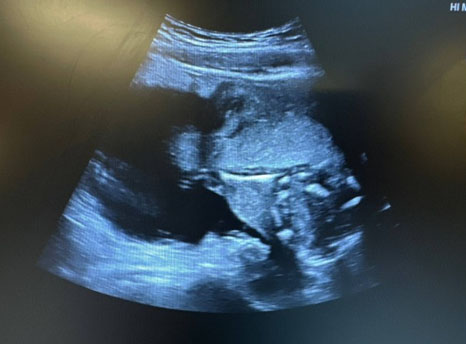
Physical examination revealed male-pattern hirsutism, clitoral hypertrophy and a painless, mobile, predominantly solid abdominal mass extending to the umbilicus (Figure 1). Laboratory evaluation revealed markedly elevated total testosterone (517 ng/dL), free testosterone (1.94 ng/dL), and androstenedione (3.9 ng/mL), with decreased sex hormone-binding globulin (SHBG) levels (15 nmol/L). Dehydroepiandrosterone sulfate (DHEA-S), 17-hydroxyprogesterone (17-OHP), cortisol, luteinizing hormone (LH), follicle-stimulating hormone (FSH), estradiol, prolactin, and other tumor markers, including CA-125, were within normal limits. Pelvic ultrasound demonstrated a right adnexal solid lesion measuring approximately 20 × 15 × 10 cm with moderate vascularization (color score 3). Magnetic resonance imaging (MRI) confirmed a large right ovarian mass without contralateral adnexal or lymph node involvement (Figure 2). The patient underwent right adnexectomy with omentectomy and peritoneal cytological analysis via laparotomy. Macroscopically, the right adnexa weighed 2880 g and comprised a normal fallopian tube and an enlarged ovary measuring 198 × 147 × 97 mm, with a smooth external surface and mixed contents. Microscopically, the tumor showed heterogeneous architecture with variable cellularity. Sheets of polygonal cells with abundant eosinophilic cytoplasm were present, corresponding to Leydig cells. No lymphovascular invasion was identified, and peritoneal fluid cytology showed no abnormal cells. The final diagnosis was a moderately differentiated SLCT, FIGO stage IA. Genetic analysis identified two pathogenic/probably pathogenic variants in the DICER1 gene; both alterations were confirmed to be non-germline.
The tumor was staged as FIGO IA, and surveillance strategy was adopted. At the 6-month postoperative reassessment, total testosterone and the remaining hormonal parameters had returned to normal levels. The clinical manifestations of hyperandrogenism, including hirsutism and virilization, had completely resolved. The patient remained under clinical surveillance for more than three years after surgery, with no evidence of disease recurrence.


Discussion
Sertoli–Leydig cell tumors are rare ovarian sex cord–stromal neoplasms that predominantly affect young women and may present with androgenic manifestations. Virilization, particularly when progressive or accompanied by markedly elevated testosterone, should prompt consideration of an androgen-secreting ovarian tumor. In the present case, the combination of rapidly progressive hirsutism, clitoromegaly, a large unilateral ovarian mass and marked testosterone elevation was highly suggestive of a hormonally active ovarian neoplasm. The normal DHEA-S, 17-hydroxyprogesterone and cortisol levels further supported an ovarian rather than adrenal source of androgen excess [2].
Imaging findings in SLCT are variable but commonly include a unilateral solid or mixed solid–cystic ovarian mass, with heterogeneous signal intensity reflecting the diverse histological components of these tumors [2],[3].
Definitive diagnosis is histopathological, with identification of the characteristic Sertoli and Leydig cell components being fundamental for tumor classification. The presence of Leydig cells provides an important morphological correlate for the androgen-secreting activity that underlies the clinical manifestations of virilization [4].
Molecular alterations involving DICER1 have emerged as an important feature of SLCT biology. Recent evidence indicates that somatic DICER1 RNase IIIb hotspot variants are particularly frequent in intermediately and poorly differentiated tumors [5],[6]. In a 2024 international registry study including 191 patients, somatic DICER1 variants were identified in 97% of intermediately and poorly differentiated tumors that underwent somatic testing [6]. The moderately differentiated morphology and somatic DICER1 alterations observed in the present case are therefore consistent with this established molecular association.
Treatment is primarily surgical, with the extent of surgery determined by disease stage, tumor differentiation, prognostic features, and the patient’s reproductive goals. Fertility-sparing surgery is an important consideration in young women with unilateral, early-stage disease when oncologically appropriate [2],[7]. In the present case, right adnexectomy with staging procedures achieved complete surgical management of FIGO stage IA disease while preserving the contralateral ovary and uterus. This approach is particularly relevant in a 23-year-old nulligravid patient.
Prognosis is generally favorable for localized SLCT, although tumor stage, degree of differentiation, and the presence of heterologous elements influence recurrence risk. In the international registry cohort, 92% of patients presented with FIGO stage I disease, and the estimated 3-year recurrence-free survival for FIGO stage IA tumors was 93.6% [6]. A national cohort similarly reported excellent cancer-related survival among patients with localized disease, with 5-year cancer-related survival of 96% for moderately differentiated tumors [8]. The absence of lymphovascular invasion, extraovarian disease and heterologous elements in the present case, together with more than three years of follow-up without documented recurrence, is consistent with the favorable outcome expected for early-stage disease.
This case highlights the importance of recognizing progressive virilization as a potential marker of an androgen-secreting ovarian neoplasm. The combination of clinical hyperandrogenism, biochemical testosterone excess, a large unilateral ovarian mass, and the subsequent identification of somatic DICER1 alterations illustrates the value of integrating clinical, radiological, pathological, and molecular findings in the diagnosis and management of SLCT.
Conclusion
Sertoli–Leydig cell tumors should be considered in young women presenting with progressive virilization and an ovarian mass. Early recognition of androgen excess may facilitate diagnosis and allow fertility-sparing treatment in selected patients with early-stage disease.
REFERENCES
1.
Young RH. Ovarian sex cord-stromal tumors: reflections on a 40-year experience with a fascinating group of tumors, including comments on the seminal observations of Robert E. Scully, MD. Arch Pathol Lab Med 2018;142(12):1459–84. [CrossRef]
[Pubmed]

2.
Muscat C, Calleja-Agius J. Review on Sertoli-Leydig cell tumours of the ovary. Discov Med 2024;36(181):234–7. [CrossRef]
[Pubmed]
3.
Cai SQ, Zhao SH, Qiang JW, Zhang GF, Wang XZ, Wang L. Ovarian Sertoli-Leydig cell tumors: MRI findings and pathological correlation. J Ovarian Res 2013;6(1):73. [CrossRef]
[Pubmed]
4.
Zhang X, Shen D, Wang Y. Detection of the DICER1 hotspot mutation alongside immunohistochemical analysis may provide a better diagnostic measure for ovarian Sertoli-Leydig cell tumors. Pathol Res Pract 2018;214(9):1370–5. [CrossRef]
[Pubmed]
5.
Schultz KAP, Harris AK, Finch M, Dehner LP, Brown JB, Gershenson DM, et al. DICER1-related Sertoli-Leydig cell tumor and gynandroblastoma: Clinical and genetic findings from the International Ovarian and Testicular Stromal Tumor Registry. Gynecol Oncol 2017;147(3):521–7. [CrossRef]
[Pubmed]
6.
Nelson AT, Harris AK, Watson D, Kamihara J, Chen KS, Stall JN, et al. Outcomes in ovarian Sertoli-Leydig cell tumor: A report from the International Pleuropulmonary Blastoma/DICER1 and Ovarian and Testicular Stromal Tumor Registries. Gynecol Oncol 2024;186:117–25. [CrossRef]
[Pubmed]
7.
Ray-Coquard I, Morice P, Lorusso D, Prat J, Oaknin A, Pautier P, et al. Non-epithelial ovarian cancer: ESMO Clinical Practice Guidelines for diagnosis, treatment and follow-up. Ann Oncol 2018;29(Suppl 4):iv1–18. [CrossRef]
[Pubmed]
8.
Bekker P, Miland-Samuelsen AR, Smerdel MP, Schnack TH, Lauszus FF, Karstensen SH. Sertoli-Leydig cell tumor: A clinicopathological analysis in a comprehensive, national cohort. Int J Gynecol Cancer 2023;33(12):1921–7. [CrossRef]
[Pubmed]
SUPPORTING INFORMATION
Acknowledgments
The authors thank the Departments of Gynecology, Radiology, Pathology of IPO-Porto and Medical Genetics for their multidisciplinary contribution.
Author ContributionsMaria Rui Torres - Conception of the work, Design of the work, Acquisition of data, Drafting the work, Final approval of the version to be published, Agree to be accountable for all aspects of the work in ensuring that questions related to the accuracy or integrity of any part of the work are appropriately investigated and resolved.
Marina Sousa Gomes - Conception of the work, Design of the work, Revising the work critically for important intellectual content, Final approval of the version to be published, Agree to be accountable for all aspects of the work in ensuring that questions related to the accuracy or integrity of any part of the work are appropriately investigated and resolved.
Monica Cruz Pires - Conception of the work, Design of the work, Revising the work critically for important intellectual content, Final approval of the version to be published, Agree to be accountable for all aspects of the work in ensuring that questions related to the accuracy or integrity of any part of the work are appropriately investigated and resolved.
Guaranter of SubmissionThe corresponding author is the guarantor of submission.
Source of SupportNone
Consent StatementWritten informed consent was obtained from the patient for publication of this article.
Data AvailabilityAll relevant data are within the paper and its Supporting Information files.
Conflict of InterestAuthors declare no conflict of interest.
Copyright© 2026 Maria Rui Torres et al. This article is distributed under the terms of Creative Commons Attribution License which permits unrestricted use, distribution and reproduction in any medium provided the original author(s) and original publisher are properly credited. Please see the copyright policy on the journal website for more information.