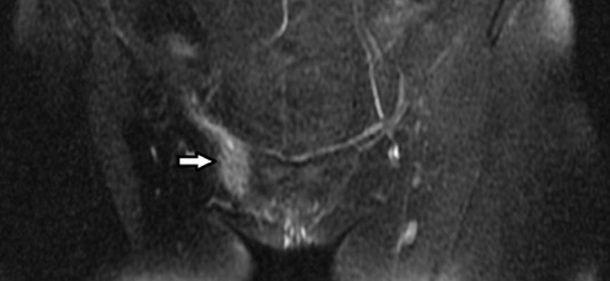
|
Case Report
Neurofibromatosis type 2 diagnosed at time of cervical squamous cell carcinoma
1 Resident, Department of Gynecologic Surgery and Obstetrics, Tripler Army Medical Center, Tripler AMC, HI, USA
2 Gynecologic Oncologist, Department of Gynecologic Surgery and Obstetrics, Tripler Army Medical Center, Tripler AMC, HI, USA
Address correspondence to:
Emily Penick
MD, FACOG, 1 Jarrett White Road, Tripler AMC, HI 96859,
USA
Message to Corresponding Author
Article ID: 100242Z08CL2026
Access full text article on other devices

Access PDF of article on other devices


How to cite this article
Lester C, Penick E. Neurofibromatosis type 2 diagnosed at time of cervical squamous cell carcinoma. J Case Rep Images Obstet Gynecol 2026;12(2):37–41.ABSTRACT
Introduction: Cervical cancer is most frequently diagnosed in women between ages 35–44 and rarely develops in younger women. Over 90% of cervical cancers are associated with human papillomavirus (HPV) persistence, but some are associated with genetic mutations such as STK11. This case discusses development of squamous cell carcinoma in a 25-year-old patient following low-grade squamous intraepithelial lesion (LSIL), cannot exclude high grade cytology result four years prior.
Case Report: A 25-year-old G2P2 presented for a postpartum exam, which was notable for a 1 cm lesion of the anterior cervix. Biopsies confirmed squamous cell carcinoma. A positron emission tomography/computed tomography (PET/CT) was completed for surgical planning which noted bilateral brain masses, concerning for vestibular schwannomas, which met clinical criteria for neurofibromatosis type 2 (NF2). Following referral to neurosurgery, a left retrosigmoid craniotomy was completed. A modified radical abdominal hysterectomy and sentinel lymph node dissection was performed following recovery from neurosurgery. Somatic genetic testing returned with a frameshift mutation in NF2E1194FJ. Final hysterectomy pathology noted a grade 3 tumor measuring 1.2 cm in size and with 3.75 mm depth of invasion.
Conclusion: Low-grade intraepithelial lesion cytology typically has a high probability of regression and low risk for progression to cancer. Neurofibromin 2 regulates many cellular events. Studies note aberrant expression of NF2 contributes to tumorigenesis in other cancers. The tumorigenesis of both HPV and NF2 may share synergistic mechanisms which lead to progression to carcinoma. While the clinical course of our patient exhibits both a clinical diagnosis of NF2 and somatic NF2 loss in her cervical tumor, we cannot definitively conclude that NF2 deficiency accelerated the timeline of her cervical carcinogenesis. Rather, this clinical scenario prompts an intriguing hypothesis regarding potential molecular synergy between high-risk HPV oncoproteins and loss of the NF2 gene product, Merlin.
Keywords: Cervical cancer, Human papillomavirus, Neurofibromatosis 2
Introduction
Cervical cancer is the fourth most common cancer among women globally and thirteenth in women in the United States [1],[2]. Annual global incidence of cervical cancer reaches approximately 660,000 individuals, with the United States seeing 13,360 of these cases (7.4–7.7 per 100,000) [1],[3],[4]. The mortality rate globally reaches around 350,000 deaths, of which 4320 occur in the United States [2],[4]. Approximately 90% of cervical cancers are attributed to the human papillomavirus (HPV) [5].
Women diagnosed with cervical cancer are rarely young. Approximately 13% of patients with cervical cancer are between the ages of 20 and 34 and the highest incidence occurs in women between 35 and 44 years (24.8%) [4]. Though regularly cleared by an individual’s immune system, HPV persistence leads to the development of cervical cancer via various oncogenic and tumorigenesis mechanisms. These mechanisms are also seen in genetic conditions such as neurofibromatosis type 2 (NF2) which itself has an extensive array of effects on cancer development. Both HPV and NF2 share pathways in regulation of p53 tumor suppressor gene and hippo signaling [6],[7],[8],[9],[10],[11],[12]. These shared pathways suggest possible synergistic mechanisms by which cervical dysplasia may progress to carcinoma. We present a case of squamous cell carcinoma in a 25-year-old female who incidentally diagnosed with NF2 at time of her cancer diagnosis.
Case Report
Informed consent was obtained from the patient for use of her case in this report. A 25-year-old G2P2 patient presented for her routine six-week postpartum visit. On pelvic exam, a 1 × 1 cm cervical mass was noted. Her gynecological history was significant for prior cervical cytology demonstrating low-grade squamous intraepithelial lesion (LSIL), cannot exclude high grade lesion. Recommendations were made by her providers to complete cervical cytology one year after the abnormal pap smear; however, this was not completed. She subsequently conceived again and additional testing was deferred at her new obstetrical visit. She went on to have a second uncomplicated vaginal delivery. She reported a history of HPV vaccination in adolescence but did not have vaccine records available to confirm dosing or timing of administration.
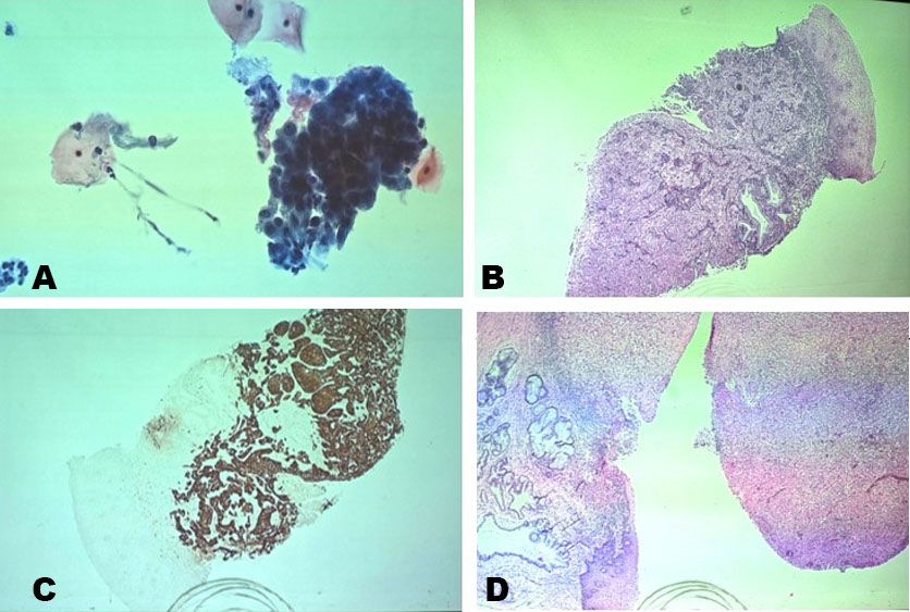
At her six-week postpartum visit following the second pregnancy, a pap smear was obtained, and the patient was referred to gynecology/oncology out of concern for a noted cervical mass. Cytology returned with atypical squamous cells of undetermined significance (ASCUS) and positive high risk-HPV testing (Figure 1A). The HPV test detected HPV types 16, 18, 31, 33, 35, 39, 45, 51, 52, 56, 58, 59, 66, 68 but was unable to specify exact HPV positive subtype.
At time of gynecologic oncology evaluation, a 1 cm exophytic, friable cervical mass with abnormal vasculature and punctuations was noted and biopsies were performed. Final pathology of the biopsies returned with invasive squamous cell carcinoma (Figure 1B and Figure 1C). A pelvic magnetic resonance imaging (MRI) and PET/CT were completed for staging and surgical planning. A comprehensive genomic and immune profiling assay, OmniSeq INSIGHT, was ordered from the colposcopy biopsies and returned with positive findings of NF2 deletion-frameshift mutation (NF2 E194fs).
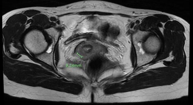
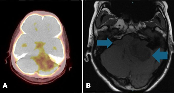
Pelvic MRI showed a sub-centimeter cervical lesion without extracervical involvement (Figure 2). Positron emission tomography/computed tomography showed increased uptake at the known cervical lesion without evidence of metastatic disease, but noted incidental findings of left brainstem and cerebellum abnormalities concerning for space occupying lesion with mass effect (Figure 3A). A primary brain lesion versus metastatic disease was suspected. Neurology was consulted and a dedicated brain MRI was ordered to further characterize the masses. Imaging noted the masses appeared consistent with bilateral vestibular schwannomas, suggestive of NF2 (Figure 3B). Significant mass effect on the pons and fourth ventricle along with mild tonsillar herniation was seen.
Neurosurgery was consulted and recommended proceeding with management of brain lesions first as there was concern the patient may develop obstructive hydrocephalus. Throughout the evaluation, the patient remained asymptomatic without neurological findings. She underwent a left retrosigmoid craniotomy for tumor resection (subtotal) within one month of the postpartum visit and pathology confirmed the masses as schwannomas, at which time she met clinical criteria for NF2. She did well postoperatively and was later followed with gynecologic oncology for surgical planning of apparent Stage IB1 squamous cell carcinoma based on clinical exam findings. Three weeks after her craniotomy she underwent a modified radical transabdominal hysterectomy, bilateral salpingectomy, bilateral sentinel lymph node mapping, ureterolysis, cystoscopy, and bilateral oophoropexy. The surgery was uncomplicated without additional findings and the patient discharged on postoperative day 1. A timeline of her care is noted in Figure 4.
Final cervical pathology demonstrated a grade 3 tumor, 1.2 cm in size, with depth of invasion to 3.75 mm, negative lymphovascular space invasion, negative margins, and negative lymph nodes (Figure 1D). Her final staging was International Federation of Gynecology and Obstetrics (FIGO) Stage IA2 based on pathology features. She did not meet Sedlis criteria for adjuvant radiation and was counseled regarding surveillance after initial recovery along with annual pap smears. She continued to receive care for vestibular schwannomas and NF2 diagnosis with the neurology team. Germline genetic testing for NF2 is pending at the time of this report.

Discussion
Low-grade intraepithelial lesion is considered low-grade cytology with a high probability for regression and low risk of progression to cancer [13]. Progression to a high-grade intraepithelial lesion (HSIL) occurs slowly in a minority of cases [6],[14],[15]. Persistent infection with high-risk HPV is a prerequisite for progression [16],[17], primarily through the oncogenic actions of the E6 and E7 proteins which disrupt key tumor suppressor pathways, including p53 and the hippo/yes associated protein (YAP) pathway, which is one of the most frequently dysregulated pathways in carcinogenesis [6],[7].
Neurofibromatosis type 2 is an autosomal dominant disorder caused by a mutation in the NF2 gene, which codes for the tumor suppressor protein Merlin [10]. Loss of Merlin function is associated with tumorigenesis which impacts a kinase cascade in the hippo pathway. The pathways disrupted by HPV appear to overlap with Merlin. For example, HPV E6 protein leads to the degradation of the p53 tumor suppressor, an effect that can also be potentiated by the loss of Merlin, which normally helps stabilize p53 [6],[10]. Similarly, both HPV E7 and loss of Merlin can lead to the activation of the oncogenic YAP/transcriptional coactivator with PDZ binding motif (TAZ) proteins in the hippo signaling pathway and promote oncogenesis [9],[11],[12].
While it is established that HPV and NF2-related pathways individually contribute to oncogenesis, it is not proven that a synergistic effect occurred in this patient. However, the concurrent diagnosis of cervical cancer and NF2 raises a compelling hypothesis that the shared disruption of the p53 and hippo/YAP pathways by both HPV and the loss of Merlin may have contributed to a more rapid clinical course than is typically expected for a young patient with a history of low-grade cytology. Further research is needed to investigate this potential interaction.
Conclusion
This case highlights the development of invasive cervical cancer in a young woman with prior low-grade cytology who was concurrently diagnosed with NF2. The shared mechanisms of p53 degradation and dysregulation of the hippo pathway describe possible synergistic mechanisms between HPV and NF2 that may have led to development of carcinoma in this 25-year-old patient. Current guidelines often recommend observation and repeat cytology in one year for low-grade cytology in patients under 25 due to high rates of regression. This case underscores the critical importance of ensuring appropriate follow-up in patients who have abnormal cervical cancer screening.
REFERENCES
1.
Cervical Cancer. World Health Organization. [Available at: https://www.who.int/news-room/fact-sheets/detail/cervical-cancer]

2.
Cancer Statistics Center. American Cancer Society. [Available at: https://cancerstatisticscenter.cancer.org/]
3.
U.S. Cancer Statistics Data Visualizations Tool. U.S. Department of Health and Human Services, Centers for Disease Control and Prevention and National Cancer Institute, released in June 2026. [Available at: www.cdc.gov/cancer/dataviz]
4.
Cancer Stat Facts: Cervical Cancer. National Cancer Institute, Surveillance, Epidemiology, and End Results Program. [Available at: https://seer.cancer.gov/statfacts/html/cervix.html]
5.
Centers for Disease Control and Prevention. Cancers Associated with Human Papillomavirus. Centers for Disease Control and Prevention, U.S. Department of Health and Human Services. 2025. [Available at: https://www.cdc.gov/united-states-cancer-statistics/publications/hpv-associated-cancers.html]
6.
Zhang Y, Qiu K, Ren J, Zhao Y, Cheng P. Roles of human papillomavirus in cancers: Oncogenic mechanisms and clinical use. Signal Transduct Target Ther 2025;10(1):44. [CrossRef]
[Pubmed]
7.
Crosbie EJ, Einstein MH, Franceschi S, Kitchener HC. Human papillomavirus and cervical cancer. Lancet 2013;382(9895):889–99. [CrossRef]
[Pubmed]
8.
Zhang H, Liu J. NF2 is a candidate diagnosis, prognostic, and immunotherapeutic biomarker: A systematic pan-cancer analysis. Transl Cancer Res 2024;13(2):1026–42. [CrossRef]
[Pubmed]
9.
Xu D, Yin S, Shu Y. NF2: An underestimated player in cancer metabolic reprogramming and tumor immunity. NPJ Precis Oncol 2024;8(1):133. [CrossRef]
[Pubmed]
10.
Kim H, Kwak NJ, Lee JY, Choi BH, Lim Y, Ko YJ, et al. Merlin neutralizes the inhibitory effect of Mdm2 on p53. J Biol Chem 2004;279(9):7812–8. [CrossRef]
[Pubmed]
11.
Fu M, Hu Y, Lan T, Guan KL, Luo T, Luo M. The Hippo signalling pathway and its implications in human health and diseases. Signal Transduct Target Ther 2022;7(1):376 [Erratum: Signal Transduct Target Ther 2024;9(1):5]. [CrossRef]
[Pubmed]
12.
Blakely WJ, Hatterschide J, White EA. HPV18 E7 inhibits LATS1 kinase and activates YAP1 by degrading PTPN14. mBio 2024;15(10):e0181124. [CrossRef]
[Pubmed]
13.
Perkins RB, Guido RS, Castle PE, Chelmow D, Einstein MH, Garcia F, et al. 2019 ASCCP risk-based management consensus guidelines for abnormal cervical cancer screening tests and cancer precursors. J Low Genit Tract Dis 2020;24(2):102–131 [Erratum: J Low Genit Tract Dis 2020;24(4):427]. [CrossRef]
[Pubmed]
14.
Bruno MT, Cassaro N, Bica F, Boemi S. Progression of CIN1/LSIL HPV persistent of the cervix: Actual progression or CIN3 coexistence. Infect Dis Obstet Gynecol 2021;2021:6627531. [CrossRef]
[Pubmed]
15.
Quint KD, de Koning MNC, Quint WGV, Pirog EC. Progression of cervical low grade squamous intraepithelial lesions: In search of prognostic biomarkers. Eur J Obstet Gynecol Reprod Biol 2013;170(2):501–6. [CrossRef]
[Pubmed]
16.
Feng T, Cheng B, Sun W, Yang Y. Outcome and associated factors of high-risk human papillomavirus infection without cervical lesions. BMC Womens Health 2023;23(1):599. [CrossRef]
[Pubmed]
17.
Schlecht NF, Platt RW, Duarte-Franco E, Costa MC, Sobrinho JP, Prado JCM, et al. Human papillomavirus infection and time to progression and regression of cervical intraepithelial neoplasia. J Natl Cancer Inst 2003;95(17):1336–43. [CrossRef]
[Pubmed]
SUPPORTING INFORMATION
Author Contributions
Clayton Lester - Conception of the work, Design of the work, Acquisition of data, Analysis of data, Drafting the work, Revising the work critically for important intellectual content, Final approval of the version to be published, Agree to be accountable for all aspects of the work in ensuring that questions related to the accuracy or integrity of any part of the work are appropriately investigated and resolved.
Emily Penick - Conception of the work, Design of the work, Acquisition of data, Analysis of data, Drafting the work, Revising the work critically for important intellectual content, Final approval of the version to be published, Agree to be accountable for all aspects of the work in ensuring that questions related to the accuracy or integrity of any part of the work are appropriately investigated and resolved.
Guaranter of SubmissionThe corresponding author is the guarantor of submission.
Source of SupportNone
Consent StatementWritten informed consent was obtained from the patient for publication of this article.
Data AvailabilityAll relevant data are within the paper and its Supporting Information files.
Conflict of InterestAuthors declare no conflict of interest.
Copyright© 2026 Clayton Lester et al. This article is distributed under the terms of Creative Commons Attribution License which permits unrestricted use, distribution and reproduction in any medium provided the original author(s) and original publisher are properly credited. Please see the copyright policy on the journal website for more information.