|
Case Report
MRI of complete hydatidiform mole with normal hCG and viable intrauterine pregnancy
1 Medical student, Technion - Israel Institute of Technology School of Medicine, Haifa, Israel
2 Department of Obstetrics and Gynecology, Rambam Health Care Campus, Haifa, Israel
3 Adjunct Professor of Radiology, The George Washington University School of Medicine, 2300 I St NW, Washington, DC 20052, USA; Chairman of Medical Imaging, Rambam Health Care Campus, HaAliya HaShniya St 8, Haifa 3109601, Israel
Address correspondence to:
Marcia C Javitt
MD, Adjunct Professor of Radiology, The George Washington University School of Medicine, 2300 I St NW, Washington, DC 20052, USA; Chairman of Medical Imaging, Rambam Health Care Campus, HaAliya HaShniya St 8, Haifa 3109601,
Israel
Message to Corresponding Author
Article ID: 100089Z08SW2021
Access full text article on other devices

Access PDF of article on other devices

How to cite this article
Weinstein S, Ginsberg Y, Reiss A, Javitt MC. MRI of complete hydatidiform mole with normal hCG and viable intrauterine pregnancy. J Case Rep Images Obstet Gynecol 2021;7:100089Z08SW2021.ABSTRACT
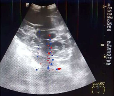
Introduction: Coexistent complete mole with a concomitant live fetus is a rare occurrence. Initially it may be identified by Ultrasound (US). Magnetic resonance imaging (MRI) is a useful alternative to US because MRI depicts the molar mass as separate from the live fetus, its monochorionic monoamniotic gestational sac and normal appearing placenta.
Case Report: A rare case of complete hydatidiform mole with concomitant live intrauterine pregnancy and normal level of serum human chorionic gonadotropin (hCG) is reported. Obstetrical US showed an indeterminate multi-cystic lenticular shaped uterine mass without ovarian theca lutein cysts. Magnetic resonance imaging showed the molar mass as separate from the live fetus within its gestational sac and a normal placenta. Although partial mole was favored because of the normal hormone level, pathology showed a complete mole after delivery of a live fetus.
Conclusion: This case highlights the utility of MRI for diagnosis and optimized management if US and clinical findings are indeterminate.
Keywords: Complete hydatidiform mole, Human serum chorionic gonadotropin, Intrauterine pregnancy
SUPPORTING INFORMATION
Author Contributions
Sarah Weinstein - Substantial contributions to conception and design, Acquisition of data, Analysis of data, Interpretation of data, Drafting the article, Revising it critically for important intellectual content, Final approval of the version to be published
Yuval Ginsberg - Analysis of data, Revising it critically for important intellectual content, Final approval of the version to be published
Ari Reiss - Analysis of data, Revising it critically for important intellectual content, Final approval of the version to be published
Marcia C Javitt - Substantial contributions to conception and design, Acquisition of data, Analysis of data, Interpretation of data, Drafting the article, Revising it critically for important intellectual content, Final approval of the version to be published
Guaranter of SubmissionThe corresponding author is the guarantor of submission.
Source of SupportNone
Consent StatementWritten informed consent was obtained from the patient for publication of this article.
Data AvailabilityAll relevant data are within the paper and its Supporting Information files.
Conflict of InterestAuthors declare no conflict of interest.
Copyright© 2021 Sarah Weinstein et al. This article is distributed under the terms of Creative Commons Attribution License which permits unrestricted use, distribution and reproduction in any medium provided the original author(s) and original publisher are properly credited. Please see the copyright policy on the journal website for more information.