 |
Case Report
Cellular angiofibroma: A rare labia minor tumor
1 Department of Obstetrics and Gynecology, Centro Hospitalar de Entre Douro e Vouga, Santa Maria da Feira, Portugal
2 Anatomical Pathology Department, Unilabs, Centro Hospitalar de Entre Douro e Vouga, Santa Maria da Feira, Portugal
Address correspondence to:
Patrícia Gomes Ferreira
Department of Obstetrics and Gynecology, Centro Hospitalar de Entre Douro e Vouga, Santa Maria da Feira,
Portugal
Message to Corresponding Author
Article ID: 100155Z08PF2023
Access full text article on other devices

Access PDF of article on other devices


How to cite this article
Ferreira PG, Carneiro C, Saraiva S, Ferreira V, Scigliano H, Monteiro I. Cellular angiofibroma: A rare labia minor tumor. J Case Rep Images Obstet Gynecol 2023;9(2):7–11.ABSTRACT
Cellular angiofibroma of the vulva is a rare benign mesenchymal tumor in middle-aged women, first reported in 1997 by Nucci et al. It is important to differentiate cellular angiofibroma from other tumors as these may be more aggressive and recurring. Cellular angiofibroma has a limited potential for local recurrence and is usually treated with complete local excision. A 45-year-old woman was referred to the gynecology appointment with a complaint of a discomfort mass in the right labium minus 1 year before, which has been progressively increasing in size for the past three months up to 6 cm. No change in vulvar skin color, local itching, or bilateral inguinal adenopathy. The tumor was excised, and the histopathological exam revealed a cellular angiofibroma. The patient recovered well and a good aesthetic result was achieved. This is the first case described of the cellular angiofibroma which arises from one of the labia minora. It is bigger in size and growing more rapidly than usual within three months. A simple excision was carried out and until now (12 months after) no recurrence signals.
Keywords: Cellular angiofibroma, Histopathology, Immunohistochemistry, Mesenchymal tumors, Vulva
Introduction
A fast-growing mass and local discomfort should be viewed as suspicious. The authors present the case of a woman with a large vulvar tumor arising from the right labium minus.
Cellular angiofibroma of the vulva is a rare benign mesenchymal tumor in middle-aged women, first reported in 1997 by Nucci et al. [1]. Mostly, it is presented in labia majora, being small-sized (<3 cm), painless, and well-circumscribed [1],[2],[3],[4]. Histopathologically, it is characterized by a spindle cell component and abundant small-sized to medium-sized thick-walled vessels, and pathologically resembles angiomyofibroblastoma and aggressive angiomyxoma [1],[5],[6]. It is important to differentiate cellular angiofibroma from other tumors as these may be more aggressive and recurring. Cellular angiofibroma has a limited potential for local recurrence and is usually treated with complete local excision [1],[2],[3],[4].
This report describes an atypical size and location of cellular angiofibroma of the vulva, which was successfully treated by excision.
Case Report
A 45-year-old woman was referred to the gynecology appointment with a complaint of a tenderness mass in the right labium minus 1 year before, which has been progressively increasing for the past three months.
The patient’s menarche was at 16 years old, and she had 5 pregnancies and eutocic deliveries. She was submitted to bilateral tubal ligation at 40 years of age. She denied any personal or family history of breast or gynecological cancer. The patient had no history of trauma, chronic infection, or insect bite.
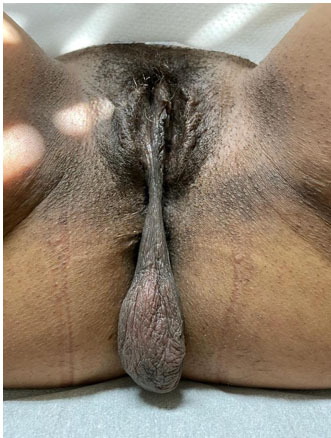
On examination, a peduncled tumor with 6 cm diameter was found, arising from the right labium minus (Figure 1), with no redness, and without signs of infection or abscess. At palpation, it was smooth and compressible. Vaginal examination was normal. No bilateral inguinal adenopathy. The patient was thoroughly investigated, and all her laboratory findings were within normal limits.
The tumor was excised with the peduncle and sent for histopathology (Figure 2). The surgical specimen was a soft grayish-brown mass measuring 6×4 cm. It was flat, well-delineated, with a disorganized fascicular pattern on section. Microscopic examination disclosed a well-circumscribed non-capsulated dermo-hypodermic benign neoplastic vascular tumor, predominantly of medium-large size. It presents a thick-walled hyalinized, arterial and venous vascular structures, with fibrous, rarely myxoid stroma, the latter showing small, monotonous, spindle cells, with bland monochromatic nuclei. Severe atypia, necrose, hemorrhage or mitosis figures were not identified (Figure 3).
Immunohistochemical study revealed positive stain for vimentin, CD34, estrogen, and progesterone receptors and absence of expression for epithelial membrane antigen (EMA) and smooth muscle actin (SMA). Overexpression of p16 nor p53 was not observed. Based on the above features, cellular angiofibroma was diagnosed (Figure 4).
Surgical excision with a free margin is the appropriate treatment for cellular angiofibroma. The postoperative period was uneventful (Figure 5). There was no recurrence in the follow-up until 12 months (Figure 6).
As there is just one case reported of recurrence and metastasis, there are no guidelines concerning the follow-up of patients with cellular angiofibroma.


Discussion
Tumors primarily arising from the vulvovaginal area are relatively rare and they include soft tissue specific and non-specific tumors, as well as a spectrum of fibro-epithelial tumors [7],[8]. Cellular angiofibroma is an uncommon benign mesenchymal neoplasm. It usually occurs on the vulva, particularly on the labia majora [9]. There are a few extra pelvic cases also reported [6].
Mandato et al. reviewed cases from 1997 to 2014 and found a total of 79 cases of female cellular angiofibroma [10]. Tumor sizes were between 0.6 and 12.3 cm, with a mean size of 3.6 cm. After this review, two vulvar cases with 20 cm were published [11],[12].
Histologically, cellular angiofibroma has two principal components: fusiform cells and prominent blood vessels. Fusiform cells form small fascicles in the middle of collagen bundles, frequent blood vessels of small to medium calibre, sometimes with a hyalinized wall, and a variable component of mature adipocytes. The tumor may present few mitotic activities and atypical cells in the stroma, but necrosis remains absent, although few instances of sarcomatous transformation and nuclear pleomorphism have also been reported [2],[4],[13],[14],[15]. The sarcomatous component can show variable features, such as: atypical lipomatous tumor, pleomorphic liposarcoma, and pleomorphic sarcoma. This phenomenon seems not to predispose to recurrence based on limited clinical follow-up available [14],[16].
All 79 cases summarized by Mandato et al. were treated with simple resection, and the surgical boundaries were not defined in 47 cases; the surgical boundary was positive in 18 cases and negative in 29 cases [10]. In the follow-up, five cases with positive surgical boundaries required re-excision [10]. In the present case, simple surgical resection was performed, and the surgical boundaries were reported to be histopathologically negative.
Of the 79 cases summarized by Mandato et al., 48 were followed up after 3–240 months; only a local recurrence of vulvar cellular angiofibroma is described after 6-month follow-up [10],[17]. Cellular angiofibroma has been initially excised with a rim of free-tumor tissue [10]. In another study of 12 patients, no recurrence or metastasis was reported at 14-month follow-up [14]. There are no reports of tumor metastasis, although the follow-up period in most of the studies is short [10],[14],[18].
Conclusion
This is the first case described of the cellular angiofibroma which arises from one of the labia minora. It is bigger sized (6 cm) than usual (<3 cm) and with rapid growth, within three months. A simple excision was carried out and until now (12 months after) no recurrence signals. In literature, a single case report of recurrence and no cases of metastasis, so there are no guidelines concerning the follow-up of patients with cellular angiofibroma.
REFERENCES
1.
Nucci MR, Granter SR, Fletcher CD. Cellular angiofibroma: A benign neoplasm distinct from angiomyofibroblastoma and spindle cell lipoma. Am J Surg Pathol 1997;21(6):636–44. [CrossRef]
[Pubmed]

2.
Colombat M, Liard-Meillon ME, De Saint-Maur P, Sevestre H, Gontier MF. Cellular angiofibroma. A rare vulvar tumor. Report of a case. [Article in French]. Ann Pathol 2001;21(2):145–8.
[Pubmed]
3.
Lane JE, Walker AN, Mullis EN Jr, Etheridge JG. Cellular angiofibroma of the vulva. Gynecol Oncol 2001;81(2):326–9. [CrossRef]
[Pubmed]
4.
Flucke U, van Krieken JHJM, Mentzel T. Cellular angiofibroma: Analysis of 25 cases emphasizing its relationship to spindle cell lipoma and mammary-type myofibroblastoma. Mod Pathol 2011;24(1):82–9. [CrossRef]
[Pubmed]
5.
Laskin WB, Fetsch JF, Mostofi FK. Angiomyofibroblastoma-like tumor of the male genital tract: Analysis of 11 cases with comparison to female angiomyofibroblastoma and spindle cell lipoma. Am J Surg Pathol 1998;22(1):6–16. [CrossRef]
[Pubmed]
6.
Iwasa Y, Fletcher CDM. Cellular angiofibroma: Clinicopathologic and immunohistochemical analysis of 51 cases. Am J Surg Pathol 2004;28(11):1426–35. [CrossRef]
[Pubmed]
7.
McCluggage WG. Recent developments in vulvovaginal pathology. Histopathology 2009;54(2):156–73. [CrossRef]
[Pubmed]
8.
Kazakov DV, Spagnolo DV, Stewart CJ, et al. Fibroadenoma and phyllodes tumors of anogenital mammary-like glands: A series of 13 neoplasms in 12 cases, including mammary-type juvenile fibroadenoma, fibroadenoma with lactation changes, and neurofibromatosis-associated pseudoangiomatous stromal hyperplasia with multinucleated giant cells. Am J Surg Pathol 2010;34(1):95–103. [CrossRef]
[Pubmed]
9.
Khmou M, Lamalmi N, Malihy A, Rouas L, Alhamany Z. Cellular angiofibroma of the vulva: A poorly known entity, a case report and literature review. BMC Clin Pathol 2016;16:8. [CrossRef]
[Pubmed]
10.
Mandato VD, Santagni S, Cavazza A, Aguzzoli L, Abrate M, La Sala GB. Cellular angiofibroma in women: A review of the literature. Diagn Pathol 2015;10:114. [CrossRef]
[Pubmed]
11.
Reder D, Kedan F, Garzon S, Haimovich S. Vulvar angiofibroma: A case report of a rare cause of huge vulvar mass. J Obstet Gynaecol 2021;41(6):990–1. [CrossRef]
[Pubmed]
12.
Ayd?n Ü, Terzi H, Turkay Ü, Eruyar AT, Kale A. A giant vulvar mass: A case study of cellular angiofibroma. Case Rep Obstet Gynecol 2016;2016:2094818. [CrossRef]
[Pubmed]
13.
Nucci MR, Fletcher CD. Vulvovaginal soft tissue tumours: Update and review. Histopathology 2000;36(2):97–108. [CrossRef]
[Pubmed]
14.
Chen E, Fletcher CDM. Cellular angiofibroma with atypia or sarcomatous transformation: Clinicopathologic analysis of 13 cases. Am J Surg Pathol 2010;34(5):707–14. [CrossRef]
[Pubmed]
15.
McCluggage WG, Ganesan R, Hirschowitz L, Rollason TP. Cellular angiofibroma and related fibromatous lesions of the vulva: Report of a series of cases with a morphological spectrum wider than previously described. Histopathology 2004;45(4):360–8. [CrossRef]
[Pubmed]
16.
Val-Bernal JF, Azueta A, Parra A, Mediavilla E, Zubillaga S. Paratesticular cellular angiofibroma with atypical (bizarre) cells: Case report and literature review. Pathol Res Pract 2013;209(6):388–92. [CrossRef]
[Pubmed]
17.
McCluggage WG, Perenyei M, Irwin ST. Recurrent cellular angiofibroma of the vulva. J Clin Pathol 2002;55(6):477–9. [CrossRef]
[Pubmed]
18.
Rodríguez-Ruiz AF, Salazar-Díaz EE, Fierro-Morales JC. Angiofibroma celular mesenquimatoso vulvar: Reporte de un caso y revisión bibliográfica. Ginecol Obstet Mex 2020;88(10):713–21.
SUPPORTING INFORMATION
Author Contributions
Patrícia Gomes Ferreira - Conception of the work, Design of the work, Acquisition of data, Analysis of data, Drafting the work, Revising the work critically for important intellectual content, Final approval of the version to be published, Agree to be accountable for all aspects of the work in ensuring that questions related to the accuracy or integrity of any part of the work are appropriately investigated and resolved.
Carolina Carneiro - Conception of the work, Design of the work, Acquisition of data, Analysis of data, Drafting the work, Revising the work critically for important intellectual content, Final approval of the version to be published, Agree to be accountable for all aspects of the work in ensuring that questions related to the accuracy or integrity of any part of the work are appropriately investigated and resolved.
Susana Saraiva - Conception of the work, Design of the work, Acquisition of data, Analysis of data, Drafting the work, Revising the work critically for important intellectual content, Final approval of the version to be published, Agree to be accountable for all aspects of the work in ensuring that questions related to the accuracy or integrity of any part of the work are appropriately investigated and resolved.
Vânia Ferreira - Conception of the work, Design of the work, Acquisition of data, Analysis of data, Drafting the work, Revising the work critically for important intellectual content, Final approval of the version to be published, Agree to be accountable for all aspects of the work in ensuring that questions related to the accuracy or integrity of any part of the work are appropriately investigated and resolved.
Horácio Scigliano - Conception of the work, Design of the work, Acquisition of data, Analysis of data, Drafting the work, Revising the work critically for important intellectual content, Final approval of the version to be published, Agree to be accountable for all aspects of the work in ensuring that questions related to the accuracy or integrity of any part of the work are appropriately investigated and resolved.
Isabel Monteiro - Conception of the work, Design of the work, Acquisition of data, Analysis of data, Drafting the work, Revising the work critically for important intellectual content, Final approval of the version to be published, Agree to be accountable for all aspects of the work in ensuring that questions related to the accuracy or integrity of any part of the work are appropriately investigated and resolved.
Guaranter of SubmissionThe corresponding author is the guarantor of submission.
Source of SupportNone
Consent StatementWritten informed consent was obtained from the patient for publication of this article.
Data AvailabilityAll relevant data are within the paper and its Supporting Information files.
Conflict of InterestAuthors declare no conflict of interest.
Copyright© 2023 Patrícia Gomes Ferreira et al. This article is distributed under the terms of Creative Commons Attribution License which permits unrestricted use, distribution and reproduction in any medium provided the original author(s) and original publisher are properly credited. Please see the copyright policy on the journal website for more information.

