|
Case Report
Spontaneous bilateral tubal ectopic pregnancy: A case report
1 MBBS, Senior House Officer, Accident and Emergency Department, Queen Elizabeth Hospital, Bridgetown, Barbados
2 Lecturer, Obstetrics & Gynaecology, Faculty of Medical Sciences, University of the West Indies, Cave Hill, Bridgetown, Barbados
Address correspondence to:
Keturah Murray
Queen Elizabeth Hospital, Martindale’s Road, St. Michael BB11155,
Barbados
Message to Corresponding Author
Article ID: 100158Z08KM2023
Access full text article on other devices

Access PDF of article on other devices


How to cite this article
Murray K, Best D. Spontaneous bilateral tubal ectopic pregnancy: A case report. J Case Rep Images Obstet Gynecol 2023;9(2):19–24.ABSTRACT
Unilateral tubal ectopic pregnancies (UTEP) are commonly encountered by physicians. Bilateral tubal ectopic pregnancies (BTEPs), however, are rare and due to the identical clinical presentations of BTEP and UTEP, there is a significant degree of diagnostic difficulty for the former, requiring a high index of suspicion. It should be considered a possibility in any newly pregnant woman of child-bearing age. The purpose of this article is to provide the reader with an example of a clinical presentation of BTEP, and a discourse on management of these patients. We present a case of a 27-year-old patient, who was seen at the emergency department of the Queen Elizabeth Hospital, Barbados, with signs and symptoms of a ruptured ectopic pregnancy. Intraoperative findings included a ruptured left ectopic pregnancy and an intact right fallopian tubal ectopic pregnancy. Left salpingectomy and right salpingostomy were performed, conserving the intact tube; her recovery was solely complicated by a superficial surgical site infection.
Keywords: Bilateral, Ectopic gestation, Ruptured, Spontaneous, Tubal, Unruptured
Introduction
Among extrauterine gestations, bilateral ectopic pregnancies are the least common [1]. They are particularly rare in the absence of assisted reproductive technologies (ART), and there have been few reported cases [2]. The clinical presentation is indistinguishable from that of a unilateral ectopic pregnancy [2]. Imaging modalities are of limited use, and the diagnosis is typically made intraoperatively, highlighting the importance of thorough examination of both fallopian tubes during the process of intraoperative evaluation of an ectopic pregnancy [3].
Case Report
A 27-year-old, Para 1+1 lady attended the emergency department of the Queen Elizabeth Hospital Barbados on August 7th, 2021, with a two-week history of generalized, sharp, non-radiating, gradually worsening lower abdominal pain, with spots of blood per vaginam and a brown vaginal discharge. She hadn’t noted any clots or fleshy material. She also reported dizziness and light-headedness, with a single syncopal episode on the day of presentation. The patient was confirmed to be sexually active, and she was unsure of the date of the first day of her last menstrual period. She had no prior pelvic or abdominal surgeries, no prior ectopic pregnancies, had not used fertility treatments and had no history of sexually transmitted infections. She had no illnesses in her family history, no drug allergies, and she did not smoke or drink alcoholic beverages.
When examined, she appeared ill, and she was hemodynamically unstable, with a blood pressure of 79/54 mmHg and pulse of 134 beats per minute (bpm). Her 12-lead electrocardiogram (ECG) demonstrated a sinus tachycardia of 124 bpm. Examination further revealed pale mucous membranes. Her abdomen was visibly distended, and on palpation exhibited generalized tenderness and guarding, but no rebound. On vaginal examination, mild cervical motion tenderness was elicited on the left. There was no bleeding, fleshy material or clots visualized on speculum examination.
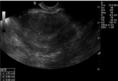
A bedside transabdominal ultrasound revealed free fluid in the pelvis, and no evidence of an intrauterine gestation. The uterus did not appear significantly enlarged, and no adnexal masses were clearly visualized. Her urine pregnancy test was positive. Baseline laboratory investigations revealed a hemoglobin concentration of 7.5 g/dL and normal urea and electrolytes; however, a serum ß-hCG was not done.
It was thought at this point that the patient likely had a ruptured left ectopic pregnancy. Due to a lack of access to laparoscopic equipment, her hemodynamic instability, and the anticipation of large hemoperitoneum, her consent was gained for an emergency laparotomy and salpingectomy.
Under general anesthesia, her abdomen was opened using a Pfannenstiel incision, and a 2.2 liter hemoperitoneum was evacuated while searching for and addressing the source of hemorrhage. Her left fallopian tube and left ovary were surrounded by clots, and the fimbrial end of the left fallopian tube was seen to be bleeding. The uterus was normal in size and appearance. The right fallopian tube was distended at its ampullary region by a round mobile mass of approximately 2 cm diameter. A left salpingectomy was performed, and a decision was also taken to perform a right salpingostomy, evacuating the ectopic pregnancy from the right tube. Both specimens were sent to the hospital’s laboratory for histopathological examination.
The patient had an uneventful early postoperative period and was discharged on postoperative day three. Unfortunately, she was re-admitted to hospital two weeks later with a superficial surgical site infection, for which she received a one-week course of cefazolin and metronidazole. She was planned to have review six weeks later in the gynecology clinic; however, unfortunately, she did not attend, and was lost to follow up.
Discussion
Ruptured ectopic pregnancies represent the leading cause of maternal mortality in the first trimester [4], thus necessitating prompt intervention and appropriate management. An ectopic pregnancy is defined as the implantation of an embryo outside of the uterine cavity [5]. This is the definition generally held by authorities such as the Royal College of Obstetricians and Gynecologists. However, it is understood that the term refers to pregnancies not in the usual location, and includes cervical, cornual and interstitial ectopic pregnancies [6]. Unilateral ectopic pregnancy (UEP) is the most common form of ectopic gestation, with 95% of these located in the fallopian tube [7]. In the Caribbean region, the incidence reported in Jamaica is 1 in 28 pregnancies [8]. No rate for the country of Barbados has been published.
Bilateral tubal ectopic pregnancy (BTEP) is an extremely rare phenomenon, occurring in 1 in 725 to 1 in 1850 ectopic pregnancies [2]. Three cases were reported between 2001 and 2005 in Barbados, a country with a total population of 280,000 people [8]. Bilateral tubal ectopic pregnancy is usually the result of assisted reproductive technologies; however few cases arising from spontaneous conception have been reported [9], and this was the circumstance with the patient presented in this case, and in the three in the case series [8]. Primary bilateral ectopic pregnancy is a condition in which there are at least two concomitant natural pregnancies in the same patient, neither of which is intrauterine, with both being located in structures on opposite sides [10].
Our patient did not have any of the typical risk factors for ectopic pregnancy, in that she had no previous ectopic pregnancy, previous adnexal surgery, previous appendectomy, or history of sexually transmitted diseases [11]. Her clinical presentation however was in-keeping with the classic triad seen in patients with ectopic pregnancy, with abdominal pain, vaginal bleeding, and amenorrhea [12]. In any woman of reproductive age with a positive pregnancy test, presenting with syncope or signs of shock, a ruptured ectopic pregnancy should be suspected. Examination, as seen, may also reveal abdominal distention and marked tenderness [13]. The clinical presentation of BTEP is indistinguishable from that of UTEP [2]. Tubal rupture is common, with as many as 16% of unilateral tubal ectopic pregnancies showing signs of rupture by six weeks of gestational age [14].
A transabdominal ultrasound scan (TAS) was performed for the patient, as transvaginal ultrasonography (TVUS) was not available at the time, and this confirmed that rupture had already taken place. Transvaginal ultrasonography has revolutionized the care of ectopic pregnancies. Prior to its introduction in 1970, more than 80% of cases were recognized after rupture, with approximately 50% presenting with shock [15]. Transvaginal ultrasonography has been found to be superior to transabdominal ultrasonography in the evaluation of suspected ectopic pregnancies [16]. The use of combined TVUS and TAS provides no more information than TVUS alone [15].
Transvaginal ultrasonography affords a more detailed assessment of the ovaries and adnexal structures [17]. Almost 75% of ectopic pregnancies are identified on TVUS alone [15]. A careful and systematic approach is essential to the diagnosis of ectopic pregnancy. The sonographic visualization of an intrauterine gestational sac containing a yolk sac and/or embryo along with normal adnexal structures essentially excludes the possibility of an ectopic pregnancy [17].
Typical findings on TVUS of an ectopic pregnancy include the corpus luteum, seen as a “ring of fire” on color Doppler on the ipsilateral side in 70–85% of cases of tubal ectopic pregnancy. Approximately 60% are seen as an inhomogeneous mass or “blob sign” adjacent to the ovary and moving separately to it; 20% appear as a hyper-echoic ring or bagel sign; and 13% have an obvious gestational sac with a fetal pole, with or without fetal cardiac activity [15]. While TVUS has proven to be beneficial in the diagnosis of unilateral ectopic pregnancy, the same diagnostic certainty cannot be provided for bilateral tubal ectopic pregnancies [3]. Overall, however, a lack of TVUS in the index case, which might have been contributed to by a rush to an urgent operation, could be seen as a shortcoming, as the greater opportunity for detection of typical ultrasound features of ectopic pregnancies was lost.
The diagnosis of BTEP in this case was made intraoperatively. This has also been the situation in published literature. There has only been one case report, published by Martinez et al., where two unruptured ectopic pregnancies in the same patient were diagnosed via transvaginal ultrasound prior to surgery [18]. Definitive diagnosis is typically made intraoperatively and, depending on the gestation, confirmed by the pathologist’s report of the specimen [19]. This underscores the importance of thorough inspection of both adnexa during the interventional procedure. Early diagnostic criteria suggested by Fishback necessitated the presence of fetal and placental parts from both tubes [19]; however this was later modified by Norris who regarded the demonstration of chorionic microvilli on histopathological evaluation as sufficient to confirm the diagnosis [19]. Ectopic gestations can also be confirmed by the presence of trophoblastic tissue in the specimen [20].
A baseline serum β-hCG was not done at the time of presentation. A published review of literature performed by José et al. found that β-hCG levels do not lead to a diagnosis of primary bilateral ectopic pregnancy, and are not particularly useful [10]. In the context of her eventually having a salpingostomy, however, a baseline level might have been considered useful for monitoring purposes. Our patient was unsure of the first date of her last normal menstrual period; however, it can be noted that the typical gestational age at the time of diagnosis of BTEP is usually 7.5 weeks, ranging from 5 to 13 weeks [10].
In the featured case, a laparotomy was performed via a Pfannenstiel incision, with left salpingectomy and a right salpingostomy. Spontaneous bilateral tubal pregnancy is very rare. If there is a suspicion of BTEP, any suspicious mass must be removed, as pathological examination is needed to confirm or exclude bilateral ectopic tubal pregnancy [9]. Management is dependent on many factors including desires for future fertility, extent of tubal damage and the condition of the patient [2]. The gold standard treatment modality is laparoscopic salpingectomy or salpingostomy [21]. Surgical options include bilateral salpingectomy, bilateral salpingostomy or salpingectomy of one tube with salpingostomy of the other, with the latter having ultimately been our chosen option. Reports indicate that laparotomy, compared to laparoscopy, is the preferred route in hemodynamically unstable patients [22].
Unlike UTEP, where patients can be managed medically with methotrexate (MTX), or surgically, there are no reported cases of BTEP successfully treated primarily by MTX [21]. Ghosh et al. reported one case of spontaneous BTEP managed with MTX however this treatment failed [23]. Their patient was managed with single dose methotrexate therapy for presumed UTEP; however, after returning with an acute abdomen and syncope, she was subsequently found to have BTEP intraoperatively [23].
The patient’s postoperative course was complicated by a superficial surgical site infection (SSI). Surgical site infections (SSIs) are the most common healthcare associated infections [24]. Pathak et al. reported that gynecological surgeries (as in our patient) had a higher incidence of SSI compared to obstetric surgeries (10.3% vs 1.2% respectively), and the majority were superficial [25]. A retrospective analysis found that laparoscopic techniques offer a protective effect against SSI, odds ratio [OR], 0.28; 95% confidence interval [CI], 0.25–0.31 [26]; thus it would seem unfortunate that laparoscopic surgery could not be afforded to our patient.
The future fertility desires of the patient were not explicitly stated. This pregnancy was unplanned. The aim of salpingostomy is to increase the likelihood of a subsequent natural intrauterine pregnancy. Studies of laparoscopic salpingostomy in patients with only one tube, rates of postsurgical intrauterine pregnancy varied between 47% and 60% [10]. Future fertility rates have been found to be similar in patients who are treated surgically by laparoscopy or laparotomy [27]. No data have been published to ascertain whether these rates can be extrapolated to pertain to cases of BTEP.
Unfortunately, our patient was lost to follow-up. Persons who undergo conservative tubal surgery should be monitored closely with serial β-hCG measurements as there is a risk of persistent trophoblastic disease. Follow-up until complete resolution is deemed necessary [21]. Some patients whose β-hCG levels do not decline or persist may require further treatment with MTX or surgical intervention in symptomatic cases [28]. Persistent trophoblast rates of 3.9–11.0% after salpingostomy have been reported [6]. The National Institute for Healthcare Excellence (NICE) of the United Kingdom recommends that women undergoing salpingotomy have a serum β-hCG level taken seven days after surgery and then weekly until a negative result is obtained [6].
Due to the rarity of the condition, there are no published data outlining the recurrence rate for BTEP; however, the recurrence rate of UTEP ranges from 6% to 16% [9]. While it is widely accepted that ectopic pregnancies have a negative effect on future fertility desires, Rani et al. reported a case of a successful term pregnancy when BTEP was treated with salpingectomy for one tube and salpingostomy for the other tube [29], offering hope to our patient should she desire another child in the future.
One limitation of this case report is that the patient was lost to follow up, therefore we could not comment on the outcome of the case with regards to any further pregnancy outcomes or any subsequent complications.
The histopathologic report read as follows in Box 1:

Conclusion
Bilateral tubal ectopic pregnancy is an extremely rare occurrence with risk for significant morbidity and mortality if not identified early and managed appropriately. The primary takeaway is to underscore the importance of careful examination of both fallopian tubes intraoperatively given the limited use of typical modalities such as ultrasound in diagnosis of bilateral ectopic pregnancies.
REFERENCES
1.
Xu H. A spontaneous bilateral tubal pregnancy: A case report. Medicine (Baltimore) 2018;97(38):e12365. [CrossRef]
[Pubmed]

2.
Jena SK, Singh S, Nayak M, Das L, Senapati S. Bilateral simultaneous tubal ectopic pregnancy: A case report, review of literature and a proposed management algorithm. J Clin Diagn Res 2016;10(3):QD01–3. [CrossRef]
[Pubmed]
3.
Gathura JE, Elfeky A, McLaren R Jr, Herzog D, Grazi R. Spontaneous bilateral tubal ectopic pregnancy in a low-risk patient: A case report with implications for preoperative patient counseling. Case Rep Obstet Gynecol 2021;2021:5588869. [CrossRef]
[Pubmed]
4.
Lawani OL, Anozie OB, Ezeonu PO. Ectopic pregnancy: A life-threatening gynecological emergency. Int J Womens Health 2013;5:515–21. [CrossRef]
[Pubmed]
5.
Mummert T, Gnugnoli DM. Ectopic Pregnancy. In: StatPearls. Treasure Island (FL): StatPearls Publishing; 2023.
[Pubmed]
6.
Diagnosis and management of ectopic pregnancy: Green-top guideline No. 21. BJOG 2016;123(13):e15–55. [CrossRef]
[Pubmed]
7.
Shehata A, Namoury ME, Heider M. Rare site of ectopic pregnancy in a patient with bicornuate uterus. Case Rep Obstet Gynecol 2016;2016:6395154. [CrossRef]
[Pubmed]
8.
Robinson N, Prussia P, Chase C. A case series of spontaneous bilateral tubal pregnancy. BAMP Bulletin 2007;163:45.
9.
Hoffmann S, Abele H, Bachmann C. Spontaneous bilateral tubal ectopic pregnancy: Incidental finding during laparoscopy – Brief report and review of literature. Geburtshilfe Frauenheilkd 2016;76(4):413–6. [CrossRef]
[Pubmed]
10.
De Los Ríos JF, Castañeda JD, Miryam A. Bilateral ectopic pregnancy. J Minim Invasive Gynecol 2007;14(4):419–27. [CrossRef]
[Pubmed]
11.
Li C, Zhao WH, Zhu Q, et al. Risk factors for ectopic pregnancy: A multi-center case-control study. BMC Pregnancy Childbirth 2015;15:187. [CrossRef]
[Pubmed]
12.
Ranji GG, Rani GU, Varshini S. Ectopic pregnancy: Risk factors, clinical presentation and management. J Obstet Gynaecol India 2018;68(6):487–92. [CrossRef]
[Pubmed]
13.
Sivalingam VN, Duncan WC, Kirk E, Shephard LA, Horne AW. Diagnosis and management of ectopic pregnancy. J Fam Plann Reprod Health Care 2011;37(4):231–40. [CrossRef]
[Pubmed]
14.
Stremick KJ, Couperus K, Ashworth SW. Ruptured tubal ectopic pregnancy at fifteen weeks gestational age. Clin Pract Cases Emerg Med 2019;3(1):62–4. [CrossRef]
[Pubmed]
15.
Winder S, Reid S, Condous G. Ultrasound diagnosis of ectopic pregnancy. Australas J Ultrasound Med 2011;14(2):29–33. [CrossRef]
[Pubmed]
16.
Nahar MN, Quddus MA, Sattar A, Shirin M, Khatun A, Ahmed R, Sultana F. Comparison of transvaginal and transabdominal ultrasonography in the diagnosis of ectopic pregnancy. Bangladesh Med Res Counc Bull 2013;39(3):104–8. [CrossRef]
[Pubmed]
17.
Lee R, Dupuis C, Chen B, Smith A, Kim YH. Diagnosing ectopic pregnancy in the emergency setting. Ultrasonography 2018;37(1):78–87. [CrossRef]
[Pubmed]
18.
Martinez J, Cabistany AC, Gonzalez M, Gil O, Farrer M, Romero JA. Bilateral simultaneous ectopic pregnancy. South Med J 2009;102(10):1055–7. [CrossRef]
[Pubmed]
19.
Norris S. Bilateral simultaneous tubal pregnancy. Can Med Assoc J 1953;68(4):379–81.
[Pubmed]
20.
Dietl J, Buchholz F. Histopathology of tubal pregnancy. [Article in German]. Geburtshilfe Frauenheilkd 1986;46(11):829–30. [CrossRef]
[Pubmed]
21.
Nyakura M, Mhlanga FG, Madziyire M, Matshalaga S. Spontaneous bilateral tubal ectopic pregnancy: A case report. Pan Afr Med J 2021;38:395. [CrossRef]
[Pubmed]
22.
Sheeba M, Supriya G. Spontaneous bilateral tubal gestation: A rare case report. Case Rep Obstet Gynecol 2016;2016:8526903. [CrossRef]
[Pubmed]
23.
Ghosh A, Borlase D, Ajala T, Kelly AJ, Ibrahim Z. Dilemmas in management of bilateral ectopic pregnancies—Report of two cases and a review of current practice. Gynecol Surg 2014;11:191–6. [CrossRef]
24.
Healthcare-associated Infections: Surgical Site Infections - Annual Epidemiological Report for 2017. [Available at: https://www.ecdc.europa.eu/en/publications-data/healthcare-associated-infections-surgical-site-infections-annual-1#:~:text=The%20percentage%20of%20SSIs%20varied,the%20type%20of%20surgical%20procedure]
25.
Pathak A, Mahadik K, Swami MB, et al. Incidence and risk factors for surgical site infections in obstetric and gynecological surgeries from a teaching hospital in rural India. Antimicrob Resist Infect Control 2017;6:66. [CrossRef]
[Pubmed]
26.
Varela JE, Wilson SE, Nguyen NT. Laparoscopic surgery significantly reduces surgical-site infections compared with open surgery. Surg Endosc 2010;24(2):270–6. [CrossRef]
[Pubmed]
27.
Othman M, Karali S, Badawi K, Mossa H. Bilateral ectopic pregnancy: Case report. WebmedCentral Obstetrics and Gynaecology 2013;4(7):WMC004354. [CrossRef]
28.
Al-Quraan GA, Al-Taani MI, Nusair BM, El-Masri A, Arafat MR, Khateeb MM. Spontaneous ruptured and intact bilateral tubal ectopic pregnancy. East Mediterr Health J 2007;13(4):972–4.
[Pubmed]
29.
Rani VRS, Puliyath G. Viable intrauterine pregnancy after spontaneous bilateral tubal ectopics in a multiparous woman: A case report. J Med Case Rep 2013;7:159. [CrossRef]
[Pubmed]
SUPPORTING INFORMATION
Author Contributions
Keturah Murray - Conception of the work, Design of the work, Acquisition of data, Analysis of data, Drafting the work, Revising the work critically for important intellectual content, Final approval of the version to be published, Agree to be accountable for all aspects of the work in ensuring that questions related to the accuracy or integrity of any part of the work are appropriately investigated and resolved.
Damian Best - Conception of the work, Design of the work, Acquisition of data, Analysis of data, Drafting the work, Revising the work critically for important intellectual content, Final approval of the version to be published, Agree to be accountable for all aspects of the work in ensuring that questions related to the accuracy or integrity of any part of the work are appropriately investigated and resolved.
Guaranter of SubmissionThe corresponding author is the guarantor of submission.
Source of SupportNone
Consent StatementWritten informed consent was obtained from the patient for publication of this article.
Data AvailabilityAll relevant data are within the paper and its Supporting Information files.
Conflict of InterestAuthors declare no conflict of interest.
Copyright© 2023 Keturah Murray et al. This article is distributed under the terms of Creative Commons Attribution License which permits unrestricted use, distribution and reproduction in any medium provided the original author(s) and original publisher are properly credited. Please see the copyright policy on the journal website for more information.