
 |
Case Report
A rare occurrence of ovarian accessory tissue torsion
1 Department of Obstetrics and Gynecology, Wright State University-Boonshoft School of Medicine, Dayton, Ohio, USA
2 Department of Obstetrics and Gynecology, Wright State University, Dayton, Ohio, USA
3 Department of Obstetrics and Gynecology, Lifestages OB/GYN-Miami Valley Hospital, Dayton, Ohio, USA
Address correspondence to:
Marilyn Kindig
Department of Obstetrics and Gynecology, Wright State University, Dayton, Ohio,
USA
Message to Corresponding Author
Article ID: 100180Z08DS2024
Access full text article on other devices

Access PDF of article on other devices


How to cite this article
Schimmoeller D, Langer A, Younglove S, Emmerling S, Kindig M. A rare occurrence of ovarian accessory tissue torsion. J Case Rep Images Obstet Gynecol 2024;10(2):22–25.ABSTRACT
The presence of an accessory ovary is a rare gynecological abnormality with unclear pathophysiology. Ectopic ovarian tissue is typically discovered incidentally during laparoscopy performed for other indications; however, these ovaries can present preoperatively similar to ovarian torsion, cyst rupture, or malignancy, prompting treatment. We present the case of a 42-year-old G2P2 with a four day history of pelvic pain. Pelvic ultrasound and computed tomography (CT) revealed a presumed 4 cm ruptured ovarian cyst. Given her persistent pain and imaging findings, we proceeded with a diagnostic laparoscopy. Intraoperatively, a large amount of edematous and hemorrhagic tissue was noted involving the right fallopian tube, and a right salpingectomy was performed. Further exploration revealed a pedunculated mass originating from a twisted stalk extending from the midline posterior uterine wall into the posterior cul-de-sac. This mass was removed and sent to pathology. There were no intraoperative complications, and the patient recovered well postoperatively. Pathology report concluded that the pelvic mass represented ovarian tissue with hemorrhage. Ectopic ovaries, however rare, can present with acute pathology and should be considered in the differential diagnosis in cases of pelvic masses or pain.
Keywords: Accessory ovary, Ectopic ovary, Ovarian torsion, Pelvic pain, Torsion
Introduction
In this case, we discuss the presence of the rare gynecologic entity of ectopic ovary as well as a commonly diagnosed ovarian pathology, ovarian torsion.
The presence of an accessory ovary is a rare gynecological abnormality [1]. Several mechanisms have been suggested, including true embryonic origins secondary to abnormal migration of ovarian precursors as well as acquired origins following surgical or inflammatory changes, however, the true pathophysiology is unknown [2].
Ovarian torsion is a gynecological emergency that can occur in females of any age [3]. Torsion occurs when either a complete or partial rotation of the adnexa leads to ischemic changes. Torsion is typically associated with ovarian mass or cyst, although it can also occur in patients with elongated infundibulopelvic ligaments. Ovarian torsion can quickly lead to tissue hypoxia and necrosis, pain, and subsequent loss of ovarian function in the affected ovary, and for this reason, it is treated as a surgical emergency [3].
Although there have been several case reports of ectopic ovaries complicated by various pathologies, this is the first documented case report on the presence of an ectopic ovarian torsion.
Case Report
Our patient is a 41-year-old G2P2 who presented to the emergency department (ED) with acute onset pelvic pain that began abruptly during sexual intercourse. In the emergency department, she was found to have a presumed 4 cm right ovarian cyst with rupture, which was visualized on both pelvic ultrasound and CT imaging. Following her diagnosis, the patient was discharged from the emergency department with narcotics. However, she presented to the emergency department again four days later with worsening pain.
The patient had a past medical history significant for irritable bowel syndrome and a history of an abnormal cervical Papanicolaou test showing mild cervical dysplasia. She had a past surgical history of laparoscopic appendectomy.
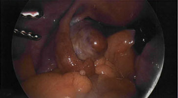
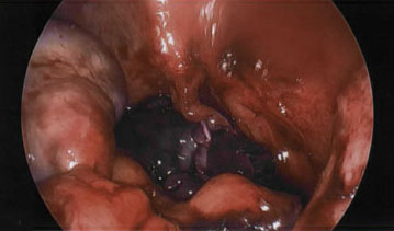
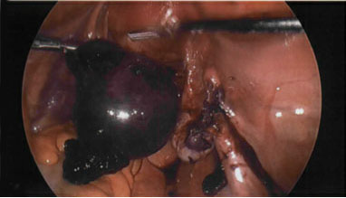
Given imaging findings on pelvic ultrasound and CT scan in addition to worsening pain, the decision was made to proceed with diagnostic laparoscopy. Upon entering the pelvis, the right fallopian tube was identified, and a large amount of edematous and hemorrhagic tissue was noted. Salpingectomy was performed. Further exploration revealed hemorrhagic tissue and debris in the posterior cul-de-sac as well as a frank blood clot. The right and left normal appearing ovaries were seen, attached to the ovarian ligament in their normal position (Figure 1 and Figure 2). With further examination, a large pedunculated mass was visualized arising from a thin twisted stalk that originated from the posterior uterine wall at the midline (Figure 1, Figure 3, and Figure 4). A bipolar diathermy (LigaSure) device was used to transect the stalk and remove the mass from the pelvis. Once removed, the mass was further examined and noted to have both solid and cystic components as well as an overall hemorrhagic appearance. The specimen was sent to pathology. Otherwise, the laparoscopic surgical procedure was uneventful. The patient had an uneventful recovery.
The histopathology report showed ovarian tissue with hemorrhage, vascular congestion, and acute inflammation as well as a full cross section of fallopian tube with areas of acute inflammation.

Discussion
The occurrence of an ectopic ovary is a rare gynecological abnormality. Though several case reports describe findings of ectopic ovaries, there is no concise incidence of the condition cited in current literature.
Although there is no clear classification system or definitive etiology for ectopic ovaries, most researchers agree that ectopic ovarian tissue can be divided into one of two categories: true embryonic and acquired. A true embryonic ectopic ovary is derived from a duplication in the genital ridge and Mullerian duct or an abnormality in the development or migration of the embryonic ovary [2],[4] . In cases where true embryonic ectopic ovarian tissue is present, additional abnormalities in the genitourinary tract is also common [5]. An acquired ectopic ovary is any ectopic ovarian tissue that arises after the descent of the fetal ovary into the pelvis. Acquired ectopic ovaries are typically associated with post-surgical or post-inflammatory changes and includes two sub-categories, the accessory and the supernumerary ovary [5]. An accessory ovary is tissue that exists in close proximity to a normal ovary and can even be connected to the normal ovary. A supernumerary ovary, on the other hand, exists separate to the normal ovaries and without any connections to the normal ovaries or pelvic ligaments [6].
There is no way to confidently classify our case of ectopic ovarian torsion. However, due to the presence of both ovarian tissue as well as fallopian tube, we speculate that this ectopic ovary was either a supernumerary ovary or a true embryonic ectopic ovary.
Conclusion
The presence of an ectopic ovary is a rare occurrence. There are few documented cases in literature, possibly due to their benign nature leading to an overall low detection rate. If discovered, these anomalies are often found incidentally on imaging or during an unrelated abdominal or pelvic surgery. Due to their presence in unexpected locations and potential interference with normal anatomy, surgeons need to keep in mind that an ectopic ovary may require adjustments be made throughout planned cases. On rare occasions, ectopic ovaries present with pathology themselves, as seen in this case. Clinicians should be aware of the remote potential for ectopic ovarian pathology and consider the possibility of their presence in female patients with unusual pelvic symptoms.
REFERENCES
1.
Muckle CW, Feinberg EC. Developmental abnormalities of the female reproductive organs. Glob Libr Women's Med 2008. [CrossRef]

2.
Nishio E, Hirota Y, Yasue A, Nishizawa H, Tsukada K, Udagawa Y. Two cases of ectopic ovary and one case of potential ectopic ovary. Reprod Med Biol 2010;10(1):51–4. [CrossRef]
[Pubmed]
3.
Huang C, Hong MK, Ding DC. A review of ovary torsion. Ci Ji Yi Xue Za Zhi 2017;29(3):143–7. [CrossRef]
[Pubmed]
4.
Pan J, Wang S, Wang H, Fan Z. A rare ectopic ovary mimicking colon sigmoideum mesenchymoma presenting as an intestinal mesenchymoma. Front Oncol 2019;9:580. [CrossRef]
[Pubmed]
5.
Kollia P, Kounoudes C, Veloudis G, Giannakou N, Gourgiotis S. True ectopic ovary in the right iliac fossa mimicking acute appendicitis and associated with ipsilateral renal agenesis. J Obstet Gynaecol Res 2014;40(3):858–61. [CrossRef]
[Pubmed]
6.
Litos MG, Furara S, Chin K. Supernumerary ovary: A case report and literature review. J Obstet Gynaecol 2003;23(3):325–7. [CrossRef]
[Pubmed]
SUPPORTING INFORMATION
Author Contributions
Daniele Schimmoeller - Analysis of data, Drafting the work, Revising the work critically for important intellectual content, Final approval of the version to be published, Agree to be accountable for all aspects of the work in ensuring that questions related to the accuracy or integrity of any part of the work are appropriately investigated and resolved.
Adam Langer - Conception of the work, Design of the work, Acquisition of data, Analysis of data, Drafting the work, Revising the work critically for important intellectual content, Final approval of the version to be published, Agree to be accountable for all aspects of the work in ensuring that questions related to the accuracy or integrity of any part of the work are appropriately investigated and resolved.
Samantha Younglove - Conception of the work, Design of the work, Acquisition of data, Analysis of data, Drafting the work, Final approval of the version to be published, Agree to be accountable for all aspects of the work in ensuring that questions related to the accuracy or integrity of any part of the work are appropriately investigated and resolved.
Susan Emmerling - Conception of the work, Design of the work, Acquisition of data, Analysis of data, Drafting the work, Final approval of the version to be published, Agree to be accountable for all aspects of the work in ensuring that questions related to the accuracy or integrity of any part of the work are appropriately investigated and resolved.
Marilyn Kindig - Analysis of data, Drafting the work, Revising the work critically for important intellectual content, Final approval of the version to be published, Agree to be accountable for all aspects of the work in ensuring that questions related to the accuracy or integrity of any part of the work are appropriately investigated and resolved.
Guaranter of SubmissionThe corresponding author is the guarantor of submission.
Source of SupportNone
Consent StatementWritten informed consent was obtained from the patient for publication of this article.
Data AvailabilityAll relevant data are within the paper and its Supporting Information files.
Conflict of InterestAuthors declare no conflict of interest.
Copyright© 2024 Adam Langer et al. This article is distributed under the terms of Creative Commons Attribution License which permits unrestricted use, distribution and reproduction in any medium provided the original author(s) and original publisher are properly credited. Please see the copyright policy on the journal website for more information.


