|
Case Series
Flexible cystoscopy and vaginoscopy: A case series on assisted endometrial biopsy to overcome anatomic barriers
1 Department of Obstetrics & Gynecology, Jersey City Medical Center, 355 Grand Street, Jersey City, NJ 07302, USA
Address correspondence to:
Alexa L Walsh
Department of Ob-stetrics & Gynecology, Jersey City Medical Center, 355 Grand Street, Jersey City, NJ 07302,
USA
Message to Corresponding Author
Article ID: 100203Z08AW2025
Access full text article on other devices

Access PDF of article on other devices


How to cite this article
Walsh AL, Salmen N, Bruck L. Flexible cystoscopy and vaginoscopy: A case series on assisted endometrial biopsy to overcome anatomic barriers. J Case Rep Images Obstet Gynecol 2025;11(1):73–76.ABSTRACT
This case series explores an innovative technique for endometrial sampling in patients facing anatomical challenges, such as cervical stenosis and morbid obesity, which often hinder traditional methods. Endometrial sampling is essential in diagnosing abnormal uterine bleeding, yet limitations such as patient tolerance, inadequate tissue procurement, and anatomical barriers can complicate the procedure. Existing solutions, including chemically induced cervical dilation and endocervical resection, pose additional risks. The report highlights two clinical cases of successful endometrial biopsies achieved using a flexible cystoscope. The first patient, a morbidly obese 35-year-old with severe cervical stenosis, underwent an examination under anesthesia and attempted hysteroscopic dilation and curettage, ultimately achieving success on the second attempt with the cystoscope. The second case involved a 77-year-old, never sexually active patient who also faced cervical stenosis, but was successfully biopsied with the flexible cystoscope after initial challenges. This technique, which enhances access to the endometrial cavity without introducing new risks, represents a promising alternative for patients with complex anatomical barriers, contributing valuable insights to the existing literature on endometrial sampling procedures. Learning point: the use of a flexible cystoscope can successfully facilitate endometrial sampling in patients with anatomical barriers such as cervical stenosis and morbid obesity, offering a safer alternative to traditional methods.
Keywords: Abnormal uterine bleeding, Anatomical barriers, Case report, Cervical stenosis, Endometrial biopsy, Flexible cystoscopy, Hysteroscopy, Uterine pathology, Vaginoscopy
Introduction
The gold standard for evaluating intrauterine pathology is direct sampling of the endometrial tissue [1]. Sampling can be done via outpatient endometrial biopsy or via a same day operative procedure of dilation and curettage (blind or under direct visualization via hysteroscopy). Adequate tissue sampling, by any method, can be limited by age, menopausal status, genitourinary anatomy, and body habitus. Limitations in patient anatomy can lead to procedural barriers such as difficulty entering the vaginal canal with the necessary instruments, visualization of the cervix, and/or inability to traverse the cervical ostium. The use of alternate methods of entry into the endometrial cavity is imperative in the workup of intrauterine pathology. In this report, we will discuss two cases of anatomical barriers preventing traditional methods of endometrial sampling and the use of a flexible cystoscope in overcoming these barriers.
Case Series
Case 1
Investigation (Patient A)
A 35-year-old gravida 0 initially presented to her primary OBGYN for abnormal uterine bleeding. The patient had been experiencing ongoing vaginal bleeding for the past seven months that was accompanied by passage of blood clots. The patient’s medical history was significant for class III obesity [body mass index (BMI) 115.52 kg/m2], chronic hypertension, depression, and asthma.
Diagnosis
Evaluation of her vaginal bleeding included a transvaginal and transabdominal ultrasound; however, the transvaginal ultrasound was unable to be performed due to body habitus. The transabdominal ultrasound revealed a uterus size of 11.1 × 5.2 × 6.0 cm with an endometrial thickness of 16 mm. The patient was subsequently referred to the gynecologic oncologist for further workup.
The patient’s initial workup through the gynecologic oncology department consisted of a pap smear (negative for intraepithelial malignancy) and an unsuccessful outpatient endometrial biopsy that showed only endocervical tissue. The decision was made to proceed with hysteroscopy dilation and curettage for sampling. Exam under anesthesia revealed a 1 cm urethral mass and a stenotic cervical os that was impenetrable to the traditionally used Hank dilators, uterine sound, endometrial pipelle, and uterine explora. The procedure was aborted.
Treatment
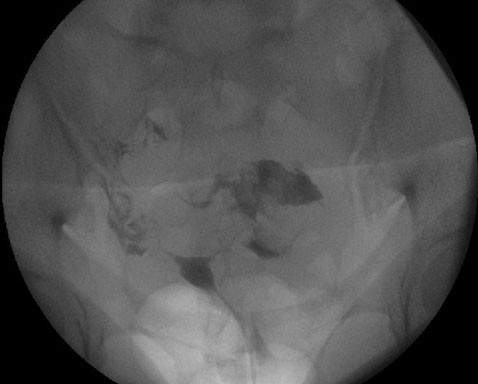
The patient was further scheduled for cystoscopy with bladder and urethral biopsies to assess the urethral mass seen on prior hysteroscopy. The gynecologic oncology team accompanied urology to attempt another hysteroscopy. With the patient’s history of cervical stenosis, the decision was made to attempt hysteroscopy using the flexible cystoscope. A 16 French flexible cystoscopy was used to first perform vaginoscopy and then easily traversed the cervical canal and allowed direct visualization of the endometrial cavity. Intrauterine survey revealed a large polypoid mass that was biopsied using a gastroenterology snare. Per chart review, the snare was able to collect an adequate sample and there were no long-term complications reported in subsequent gynecologic follow-up.
Case 2
Investigation (Patient B)
A 77-year-old virginal gravida 0 was referred to the gynecologic oncology clinic for endometrial biopsy after a transvaginal ultrasound demonstrated an endometrial thickness measuring 10 mm. The patient had never experienced postmenopausal bleeding; therefore, she was given the option of closer observation versus diagnostic management. The patient opted for diagnostic measures and the decision was made to proceed with exam and endometrial biopsy under anesthesia.
Diagnosis
Exam under anesthesia revealed a vaginal introitus with a diameter of 1 cm that inhibited the insertion of a speculum or retractors. The cervix was palpated using one digit and found to be small and firm. A 5 mm hysteroscope was then introduced to visualize the cervix, but biopsy using an endometrial pipelle was unsuccessful. The 16 French flexible cystoscope was then used to perform vaginoscopy and successfully used to traverse the cervical os. The endometrial biopsy was again attempted using an endometrial pipelle with success. Samples were then sent to pathology. Per chart review, the samples obtained were adequate for diagnosis and there were no long-term complications reported in subsequent gynecologic follow-up.
Discussion
Endometrial biopsy can be performed in either the outpatient or inpatient setting and is referred to as a relatively quick and cost-effective diagnostic procedure in the workup of intrauterine pathology [2]. Endometrial sampling was found to be 80.56% successful (via endometrial pipelle) and 91.11% successful (via dilation and curettage) [3]; however, women with anatomical barriers such as a narrow vaginal introitus or severely stenotic cervical os are unable to reap the diagnostic benefits. Traditional means of mechanical cervical dilation in the setting of stenotic cervix carry the risk of uterine perforation and its sequelae [4]. More recent studies have discussed the use of chemical dilation using hyoscine butyl bromide or hysteroscopic endocervical excision to aid in intrauterine access [4],[5],[6]. Studies investigating the use of hyoscine butyl bromide noted its effectiveness in cervical dilation in pre-menopausal women prior to intrauterine procedure; however, several potential side effects were addressed. These side effects included local skin reaction, tachycardia, dry mouth, urinary retention, and gastric irritation [7]. Investigation regarding cervical excisional procedures to aid in biopsy, again, show useful in entering the cervix, but are not favored in women desiring fertility due to the effects on cervical structure [5]. Other studies discussed the possibility of entry into the descending branches of the uterine vessels, which may result in greater blood loss and longer time under anesthesia [5]. Treatment with the flexible cystoscopy as described above did not show similar adverse effects in our patients. Additional research is necessary to compare the use of flexible cystoscopy versus chemical dilators and excisional procedures.
There are no recent reports of cystoscope use to aid in endometrial biopsy. Our technique using the flexible cystoscope in two previously mentioned cases combines the aspect of using a singular, small instrument capable of entering a stenotic cervix and the aspect of performing this under direct visualization. This technique provides a means of overcoming various anatomical barriers without additional procedures, medications, or alterations in the patient’s anatomy while permitting adequate endometrial sampling. There have been no comparative studies regarding the use of medication or excisional procedure versus flexible cystoscope to achieve successful endometrial biopsy. Future studies research comparing the outcomes and adverse reactions of these methods.
Conclusion
- Importance of Patient-Specific Considerations: Both cases highlight how individual patient factors, such as age, body habitus, and anatomical abnormalities, can complicate standard procedures for endometrial sampling. This underscores the necessity for healthcare providers to assess and adapt to the unique anatomical and health characteristics of each patient to optimize diagnostic outcomes.
- Advancement of Techniques for Challenging Anatomy: The successful use of flexible cystoscopy to facilitate endometrial biopsy in both cases illustrates an important advancement in technique. This method was adapted to overcome procedural barriers that typically hinder traditional approaches. This suggests that healthcare providers should be knowledgeable about alternative methods and tools that can be employed in similar situations.
- Collaborative Multidisciplinary Approach: Both cases demonstrate the benefits of a collaborative approach between gynecologists and urologists to ensure comprehensive patient care. This approach can lead to more effective management strategies and ultimately better diagnostic accuracy.
REFERENCES
1.
Ozelci R, Dilbaz B, Akpınar F, Kınay T, Baser E, Aldemir O, Altınbas SK. The significance of sonographically thickened endometrium in asymptomatic postmenopausal women. Obstet Gynecol Sci 2019;62(4):273–9. [CrossRef]
[Pubmed]

2.
Will AJ, Sanchack KE. Endometrial Biopsy. In: StatPearls. Treasure Island (FL): StatPearls Publishing; 2025.
3.
Tanko NM, Linkov F, Bapayeva G, et al. Pipelle endometrial biopsy for abnormal uterine bleeding in daily clinical practice: Why the approach to patients should be personalized? J Pers Med 2021;11(10):970. [CrossRef]
[Pubmed]
4.
Hadadianpour S, Tavana S, Tavana A, Fallahian M. Immediate dilation of a tight or stenotic cervix by intra-procedural administration of hyoscine butylbromide: A clinical trial. Int J Reprod Biomed 2019;17(4):253–60. [CrossRef]
[Pubmed]
5.
Suen MWH, Bougie O, Singh SS. Hysteroscopic management of a stenotic cervix. Fertil Steril 2017;107(6):e19. [CrossRef]
[Pubmed]
6.
Wortman M, Daggett A. Hysteroscopic endocervical resection. J Am Assoc Gynecol Laparosc 1996;4(1):63–8. [CrossRef]
[Pubmed]
7.
Hadadian S, Fallahian M. Assessing the efficacy of vaginal hyoscine butyl bromide on cervical ripening prior to intrauterine procedures: A double-blinded clinical trial. Int J Reprod Biomed 2016;14(11):709–12.
[Pubmed]
SUPPORTING INFORMATION
Acknowledgments
This research would not have been possible without the generosity and guidance provided by Dr. Nicole Salmen and Dr. Lance Bruck.
Author ContributionsAlexa L Walsh - Conception of the work, Design of the work, Acquisition of data, Analysis of data, Drafting the work, Revising the work critically for important intellectual content, Final approval of the version to be published, Agree to be accountable for all aspects of the work in ensuring that questions related to the accuracy or integrity of any part of the work are appropriately investigated and resolved.
Nicole Salmen - Conception of the work, Design of the work, Acquisition of data, Analysis of data, Drafting the work, Revising the work critically for important intellectual content, Final approval of the version to be published, Agree to be accountable for all aspects of the work in ensuring that questions related to the accuracy or integrity of any part of the work are appropriately investigated and resolved.
Lance Bruck - Conception of the work, Design of the work, Acquisition of data, Analysis of data, Drafting the work, Revising the work critically for important intellectual content, Final approval of the version to be published, Agree to be accountable for all aspects of the work in ensuring that questions related to the accuracy or integrity of any part of the work are appropriately investigated and resolved.
Guaranter of SubmissionThe corresponding author is the guarantor of submission.
Source of SupportNone
Consent StatementWritten informed consent was obtained from the patient for publication of this article.
Data AvailabilityAll relevant data are within the paper and its Supporting Information files.
Conflict of InterestAuthors declare no conflict of interest.
Copyright© 2025 Alexa L Walsh. This article is distributed under the terms of Creative Commons Attribution License which permits unrestricted use, distribution and reproduction in any medium provided the original author(s) and original publisher are properly credited. Please see the copyright policy on the journal website for more information.