|
Case Report
Retroperitoneal schwannoma: An abnormal presentation mimicking a pelvic mass
1 Department of Obstetrics and Gynecology, Medical City Arlington, 3301 Matlock Road, Arlington, TX, USA
2 Texas College of Osteopathic Medicine, 3500 Camp Bowie Boulevard, Fort Worth, TX, USA
Address correspondence to:
Isabella Sciacca
3201 Matlock Road, Suite 220, Arlington, TX 76015,
USA
Message to Corresponding Author
Article ID: 100212Z08IS2025
Access full text article on other devices

Access PDF of article on other devices


How to cite this article
Sciacca I, Reeves C, Stokes C, Seligman S. Retroperitoneal schwannoma: An abnormal presentation mimicking a pelvic mass. J Case Rep Images Obstet Gynecol 2025;11(2):18–22.ABSTRACT
Introduction: A schwannoma is an encapsulated nerve sheath tumor originating from differentiated Schwann cells. Schwannomas are preferentially localized to the head, neck, and extremities with a minor occurrence of 1–3% in the retroperitoneal space. Schwannomas are often asymptomatic due to slow growth but can be present with symptoms according to their location. We report a case of a 45-year-old female with a retroperitoneal schwannoma that initially was suspected to be an ovarian mass.
Case Report: A 45-year-old female with a 10-year history of a right ovarian cyst presented with lower abdominal pain, early satiety, hematuria, low back pain and constipation. Transabdominal ultrasound showed right ovarian enlargement measuring 8.8 × 6.6 cm, described as heterogeneous with loculations. Tumor markers indicated a low risk of malignancy. The patient was taken to the operating room for an exploratory laparotomy that revealed a large, firm posterior pelvic mass palpated in the retroperitoneal space. The cyst wall could not be completely dissected due to proximity to major vessels and nerve roots. The patient was referred to a neighboring academic institution because of the case complexity. She underwent repeat exploratory laparotomy and neuroplasty of the L5, S1, and S2 nerve roots. The L5 nerve root was ultimately sacrificed and the retroperitoneal mass was removed. Final pathology revealed a benign schwannoma.
Conclusion: The diagnosis of a retroperitoneal schwannoma is often missed because of its rare occurrence, location, vague symptoms, and resemblance to other tumors on varying imaging modalities. Due to low incidence and often delay in diagnosis, current management guidelines are limited. Currently, histopathological exam after total surgical resection is the most reliable source for diagnosis. The patient in this case demonstrates the need for improvement in the preoperative evaluation and consideration of non-gynecologic differential diagnoses in the evaluation of a suspected pelvic mass.
Keywords: Case report, Misdiagnosis, Ovarian mass, Pelvic mass, Retroperitoneal schwannoma, Retroperitoneum, Schwannoma
Introduction
A schwannoma is an encapsulated nerve sheath tumor originating from differentiated Schwann cells of the peripheral nervous system derived from the neural crest [1]. The majority of schwannomas are sporadic in nature, with a small portion related to neurofibromatosis type 2 (NF2), schwannomatosis or Carney’s complex [2]. Most cases are benign, with malignant transformation exceedingly rare [3]. They are preferentially localized to the head, neck, and extremities [4]. Schwannomas rarely occur in the retroperitoneal space, accounting for only 1–3% of all schwannomas [5]. Of the tumors appearing within the retroperitoneal space, schwannomas account for only 4% [5]. Presenting symptoms specific to the retroperitoneal space are typically vague abdominal complaints such as abdominal discomfort, with complaints of flank pain, hematuria, and hypertension being less common [6]. As a result of the non-restrictive nature of the retroperitoneum, tumors can grow to be large prior to causing symptoms [7]. We report a case of a 45-year-old female with a retroperitoneal schwannoma that initially was suspected to be an ovarian mass.
Case Report
We report the case of a 45-year-old premenopausal female with a past medical history of endometriosis, in vitro fertilization (IVF), substance use disorder, and a right ovarian cyst first diagnosed over 10 years ago. Upon presentation, she reported three weeks of sharp lower abdominal pain, early satiety, fatigue, decreased appetite, nausea, chills, hematuria, lower back pain, and constipation. In the three weeks prior to her presentation, she visited an urgent care clinic multiple times due to the previously mentioned symptoms. At the urgent care clinic, she was found to have right ovarian enlargement (7.7 × 7.1 × 8.5 cm) on ultrasound with an associated mass described to have multifocal cystic lesions and septations. She had reportedly seen multiple physicians since 2013 who had recommended surgical removal of the mass, but the patient was lost to follow-up.
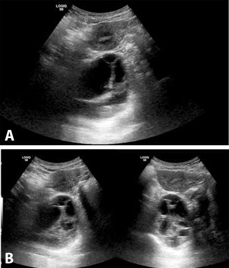
When she presented, transvaginal ultrasound showed right ovarian enlargement measuring 8.8 × 6.6 cm, described as heterogeneous with loculations (Figure 1). There was no evidence of ascites or obvious structures on the capsule of the ovary. CA 125 was 12.8 (reference range: 0.0–38.1). HE4 was 43.4 (reference range: 0.0–63.6). Risk for Ovarian Malignancy (ROMA) score was 0.54, indicating a low likelihood of finding a malignancy on surgery.
Due to the low risk of malignancy and suspected intermittent ovarian torsion, the patient was counseled and agreed to surgical management with a diagnostic laparoscopy with the possibility of exploratory laparotomy, right salpingo-oophorectomy with possible total abdominal hysterectomy. An exam under anesthesia revealed a fixed mass to the right pelvic side wall. The decision was made for exploratory laparotomy due to the anticipated difficulty dissection and the surgeon’s desire for digital dissection of the mass to facilitate removal. Exploratory laparotomy was performed through a Pfannenstiel incision due to the patient’s history of a prior cesarean section. On visualization, the uterus, fallopian tubes, and ovaries were normal appearing without masses. A large, firm, posterior pelvic mass was palpated in the retroperitoneal space with the right ureter transversing superior to the mass. The retroperitoneum was entered, and the ureter was dissected free. Following this the firm cystic mass was separated from the surrounding capsule and inadvertently drained. The fluid was serous, and the inside of the mass had areas of what appeared to be caseating necrosis and complex in nature. The cyst wall could not be fully dissected due to adherence of the mass to the iliac vessels. Total abdominal hysterectomy was not performed.
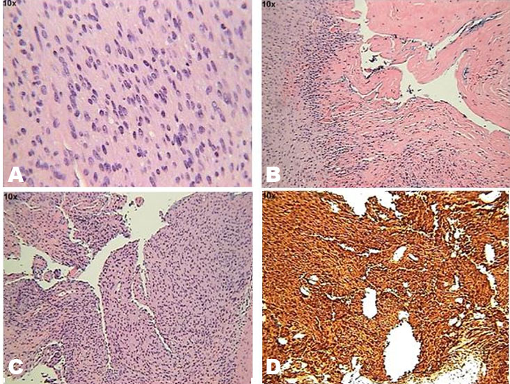
The pathology of the retroperitoneal mass showed a spindle cell neoplasm with fibrous encapsulation and cellular areas with mild nuclear pleomorphism, representing Antoni A areas (Figure 2). Cystic areas representing Antoni B degeneration were also visualized (Figure 2). The tumor was strongly positive for S100 protein in the spindle cell population (Figure 2). Final diagnosis of a right retroperitoneal schwannoma without evidence of malignancy was reported.
On post-operative day 2, the patient reported subjective fevers 3–4 times daily, right flank pain and sciatica down her right leg. Computed tomography (CT) of the abdomen/pelvis showed mild dilation of the right collecting system and right ureter with a large heterogeneous focus along the right adnexa measuring 8.5 × 9.4 × 8.9 cm. This focus showed a focal region of air and fluid along the anterior margin measuring 1.8 × 4.2 × 2.8 cm with surrounding stranding and inflammation extending along the lower abdomen and pelvis. Urology was consulted and performed a cystoscopy, bilateral retrograde pyelogram and right ureteral stent insertion prior to discharge from the hospital.
The patient was then referred to a neighboring institution for a higher level of care for further management of her benign schwannoma. On initial examination with surgical oncology, the patient complained of spine and right lower extremity pain. The patient was referred to orthopedic oncology due to concern for nerve involvement. She was found to have L4, L5, and S1 radiculopathy. Electromyography (EMG) showed evidence of right S1 nerve root degeneration. A combined surgical case between surgical oncology and orthopedic oncology was planned. She underwent exploratory laparotomy with extensive lysis of adhesions, right salpingo-oophorectomy, removal of right pelvic mass and neuroplasty of the L5, S1, and S2 nerve roots. Intraoperative findings included the ureter encased in scar tissue and adhered to the pelvic mass. Additionally, the pelvic mass was noted to be adhered to the L5, S1, and S2 nerve roots causing a compression effect and displacing the internal and external iliac vessels. The mass was intimately attached to the L5 nerve root with impressive splaying of the nerve directly into the mass, necessitating the L5 nerve fibers to be sacrificed. This intimate attachment demonstrated the progression of the patient’s schwannoma as a result of years of non-treatment and misdiagnosis. The pelvic mass was removed in its entirety and sent to pathology.
Pathology reported the specimen as a schwannoma without evidence of malignancy. Her postoperative course was complicated by permanent foot drop and chronic neuropathic pain secondary to the L5 nerve root sacrifice. The neurologic consequences following removal of this mass could possibly have been avoided given earlier suspicion of the mass being of retroperitoneal origin mitigating the complexity and morbidity of the procedure.
Discussion
The retroperitoneum is a large and flexible space that is part of the abdominal cavity that lies between the posterior parietal peritoneum anteriorly and the posterior abdominal wall [8]. While atypical, a retroperitoneal schwannoma can arise, often from the paravertebral region [9]. As a result of its rarity, location, late symptom onset and vague symptom presentation, a diagnosis of a retroperitoneal schwannoma is often missed [9]. Additionally, similarities with other tumors on varying imaging modalities can complicate the diagnostic process [10].
An undiagnosed or misdiagnosed retroperitoneal schwannoma demonstrates the necessity of ultrasound guided biopsy preoperatively; however, cellular pleomorphism in areas of degeneration can still lead to inaccurate diagnosis [10]. There is no established gold standard for diagnosing a retroperitoneal schwannoma. Currently, histopathological exam after total surgical resection is the most reliable source for diagnosis [3]. This patient was misdiagnosed for over 10 years, until complete surgical resection, due to her non-specific symptoms and reported CT, and ultrasound results that imitated a large ovarian cyst. It is possible that preoperative magnetic resonance imaging (MRI) prior to the initial surgery could have distinguished a retroperitoneal mass as opposed to an ovarian mass. Distinct MRI features such as location and mobility could have helped determine whether the mass was adnexal, typically adjacent to or attached to the ovary and mobile with uterine and ovarian movement [11]. In contrast, schwannomas are typically fixed and separate from the ovary and follow neural pathways [11]. Additionally, on MRI, schwannomas appear hypo-to-isointense on T1-weighted imaging and hyperintense on T2-weighted [12]. Schwannomas can demonstrate a target-like pattern formed by a lower T2 signal centrally with a high T2 signal peripherally [13]. This can be compared to the appearance of adnexal masses, which vary widely depending on its make up [14].
In complex cases where the diagnosis remains uncertain, it may be imperative to employ advanced imaging modalities such as MRI with neurography or positron emission tomography (PET) scans. Magnetic resonance neurography (MRN) enables detailed visualization of peripheral nerves by optimizing signal from neural water, making it particularly useful to determine whether a mass arises from or compresses nerve structures, information that may distinguish schwannomas from ovarian or adnexal tumors [15]. Positron emission tomography-computed tomography (PET-CT), while not definitive for benign versus malignant peripheral nerve sheath tumors, often reveals increased metabolic activity in schwannomas, which may mimic malignancy [16]. Nevertheless, PET can aid in assessing metabolic behavior and guiding further management when conventional imaging is inconclusive [16]. Use of these modalities could facilitate earlier and more accurate diagnosis in complex presentations, allowing timely surgical planning and potentially reducing morbidity associated with delayed treatment.
The current management guidelines of retroperitoneal schwannomas are limited. This is in part due to a low incidence and often delay in diagnosis [7]. Total surgical excision is the preferred treatment method of a retroperitoneal schwannoma [9]. Both open and laparoscopic approaches are noted to have good outcomes [3]. Incomplete excision results in recurrence in 5–10% of cases [4]. Incomplete excision is often due to adjacent nerve bundles and vessels close to the tumor that cause concern for injury, as demonstrated in this case. Due to high recurrence rates with potential incomplete excision, long term follow-up is required in retroperitoneal schwannoma treatment [3].
A benign retroperitoneal schwannoma has a good prognosis and a low likelihood of recurrence after complete surgical resection [3]. Injury to adjacent structures intraoperatively can cause significant morbidity and mortality. Careful preoperative planning, patient counseling, and consideration of non-gynecologic causes—along with early interdisciplinary consultation, such as radiology, oncology, and neurology—are imperative during the work-up of a suspected pelvic mass to improve outcomes and patient satisfaction.
Conclusion
Retroperitoneal schwannomas are rare and can present with slowly appearing vague abdominal symptoms that may mimic more common pelvic masses. This presentation leads to significant delays in diagnosis and potential complications in patient care. This case presentation highlights the importance of considering non-gynecologic causes when developing a differential diagnosis for complex pelvic masses, especially in patients with prolonged unexplained symptoms. Advanced imaging, early interdisciplinary consultation, and preoperative planning are crucial for improving a timely and accurate diagnosis to deliver adequate care and improve patient outcomes.
REFERENCES
1.
Moini J, Avgeropoulos NG, Samsam M. Chapter 10 – Schwannoma. In: Moini J, Avgeropoulos NG, Samsam M, editors. Epidemiology of brain and spinal tumors. 1st ed. Academic Press; 2021. p. 179–96. [CrossRef]

2.
Hilton DA, Hanemann CO. Schwannomas and their pathogenesis. Brain Pathol 2014;24(3):205–20. [CrossRef]
[Pubmed]
3.
Abdellatif E, Kamel D, Mantilla JG. Schwannoma. PathologyOutlines.com. [Available at: https://www.pathologyoutlines.com/topic/softtissueschwannoma.html]
4.
Debaibi M, Essid R, Sghair A, Zouari R, Sahnoun M, Dhaoui A, et al. Retroperitoneal schwannoma: Uncommon location of a benign tumor. Clin Case Rep 2022;10(4):e05726. [CrossRef]
[Pubmed]
5.
Harhar M, Ramdani A, Bouhout T, Serji B, El Harroudi T. Retroperitoneal schwannoma: Two rare case reports. Cureus 2021;13(2):e13456. [CrossRef]
[Pubmed]
6.
Cury J, Coelho RF, Srougi M. Retroperitoneal schwannoma: Case series and literature review. Clinics (Sao Paulo) 2007;62(3):359–62. [CrossRef]
[Pubmed]
7.
Holbrook C, Saleem N. Retroperitoneal schwannoma: An unusual cause of abdominal distention. BMJ Case Rep 2017;2017:bcr2017220221. [CrossRef]
[Pubmed]
8.
Jones J, Glick Y, Sharma R, et al. Retroperitoneum. Reference article, Radiopaedia.org. [Available at: https://radiopaedia.org/articles/5718]
9.
Zipple MK, Shu AL, Mahendram S, McKany ME. A case of retroperitoneal schwannoma: Determination of appropriate timing for surgical resection. ACS Case Reviews in Surgery 2020;2(6):5–8.
10.
Zhang L, Gao M, Zhang T, Chong T, Wang Z, Liu W, et al. Surgical management of retroperitoneal schwannoma complicated with severe hydronephrosis: A case report. Medicine (Baltimore) 2018;97(39):e12528. [CrossRef]
[Pubmed]
11.
Spencer JA, Ghattamaneni S. MR imaging of the sonographically indeterminate adnexal mass. Radiology 2010;256(3):677–94. [CrossRef]
[Pubmed]
12.
Rha SE, Byun JY, Jung SE, Chun HJ, Lee HG, Lee JM. Neurogenic tumors in the abdomen: Tumor types and imaging characteristics. Radiographics 2003;23(1):29–43. [CrossRef]
[Pubmed]
13.
Torigian DA, Ramchandani P, Boll D. CT and MRI of the whole body. In: Haaga JR, Boll D, editors. CT and MRI of the whole body. 6th ed. Philadelphia: Elsevier; 2017.
14.
Koç A. MRI evaluation of extremity schwannoma characteristics. CMJ 2018;40(2):115–9. [CrossRef]
15.
Rockall A, Forstner R. Adnexal diseases. In: Hodler J, Kubik-Huch RA, von Schulthess GK, editors. Diseases of the abdomen and pelvis 2018–2021: Diagnostic imaging – IDKD book [Internet]. Cham: Springer; 2018. p. 105–13. [CrossRef]
16.
Kuntz C 4th, Blake L, Britz G, Filler A, Hayes CE, Goodkin R, et al. Magnetic resonance neurography of peripheral nerve lesions in the lower extremity. Neurosurgery 1996;39(4):750–6; discussion 756–7. [CrossRef]
[Pubmed]
17.
Goh PG, Ko KH, Kim ES, Kim YJ, Lee SY, Moon HS, et al. A case of a retroperitoneal schwannoma presenting as hypermetabolic mass in PET-CT. [Article in Korean]. Korean J Gastroenterol 2011;57(5):323–6. [CrossRef]
[Pubmed]
SUPPORTING INFORMATION
Acknowledgments
We thank the Medical City Arlington Department of Pathology for assistance in obtaining and permission of use of the images in this manuscript.
Author ContributionsIsabella Sciacca - Conception of the work, Design of the work, Drafting the work, Revising the work critically for important intellectual content, Final approval of the version to be published, Agree to be accountable for all aspects of the work in ensuring that questions related to the accuracy or integrity of any part of the work are appropriately investigated and resolved.
Christelle Reeves - Conception of the work, Design of the work, Revising the work critically for important intellectual content, Final approval of the version to be published, Agree to be accountable for all aspects of the work in ensuring that questions related to the accuracy or integrity of any part of the work are appropriately investigated and resolved.
Cameron Stokes - Conception of the work, Design of the work, Drafting the work, Revising the work critically for important intellectual content, Final approval of the version to be published, Agree to be accountable for all aspects of the work in ensuring that questions related to the accuracy or integrity of any part of the work are appropriately investigated and resolved.
Steven Seligman - Conception of the work, Design of the work, Revising the work critically for important intellectual content, Final approval of the version to be published, Agree to be accountable for all aspects of the work in ensuring that questions related to the accuracy or integrity of any part of the work are appropriately investigated and resolved.
Guaranter of SubmissionThe corresponding author is the guarantor of submission.
Source of SupportNone
Consent StatementWritten informed consent was obtained from the patient for publication of this article.
Data AvailabilityAll relevant data are within the paper and its Supporting Information files.
Conflict of InterestAuthors declare no conflict of interest.
Copyright© 2025 Isabella Sciacca et al. This article is distributed under the terms of Creative Commons Attribution License which permits unrestricted use, distribution and reproduction in any medium provided the original author(s) and original publisher are properly credited. Please see the copyright policy on the journal website for more information.